

Ex-vivo cultured human corneoscleral segment model to study the effects of glaucoma factors on trabecular meshwork
- PMID: 32579557
- PMCID: PMC7314024
- DOI: 10.1371/journal.pone.0232111
Ex-vivo cultured human corneoscleral segment model to study the effects of glaucoma factors on trabecular meshwork
Erratum in
-
Correction: Ex-vivo cultured human corneoscleral segment model to study the effects of glaucoma factors on trabecular meshwork.PLoS One. 2020 Aug 25;15(8):e0238408. doi: 10.1371/journal.pone.0238408. eCollection 2020. PLoS One. 2020. PMID: 32841305 Free PMC article.
Abstract
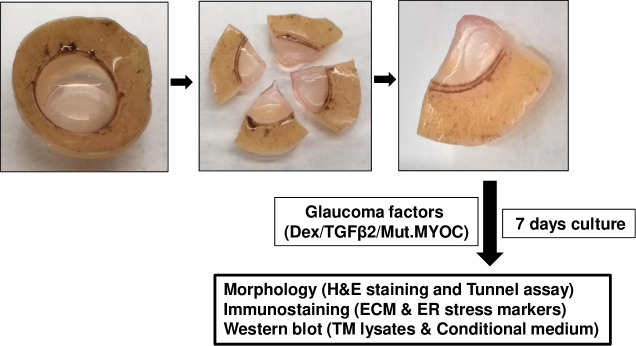
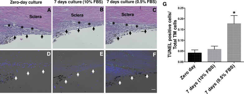
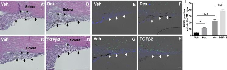
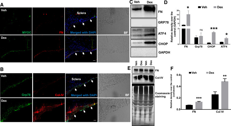
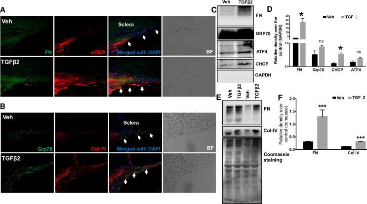
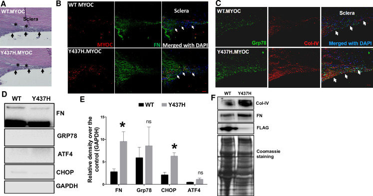
Glaucoma is the second leading cause of irreversible blindness worldwide. Primary open angle glaucoma (POAG), the most common form of glaucoma, is often associated with elevation of intraocular pressure (IOP) due to the dysfunction of trabecular meshwork (TM) tissues. Currently, an ex vivo human anterior segment perfusion cultured system is widely used to study the effects of glaucoma factors and disease modifying drugs on physiological parameters like aqueous humor (AH) dynamics and IOP homeostasis. This system requires the use of freshly enucleated intact human eyes, which are sparsely available at very high cost. In this study, we explored the feasibility of using human donor corneoscleral segments for modeling morphological and biochemical changes associated with POAG. Among the number of corneas donated each year, many are deemed ineligible for transplantation due to stringent acceptance criteria. These ineligible corneoscleral segments were obtained from the Lions Eye Bank, Tampa, Florida. Each human donor anterior corneoscleral segment was dissected into four equal quadrants and cultured for 7 days by treating with the glaucoma factors dexamethasone (Dex) or recombinant transforming growth factor (TGF) β2 or transduced with lentiviral expression vectors containing wild type (WT) and mutant myocilin. Hematoxylin and Eosin (H&E) staining analysis revealed that the TM structural integrity is maintained after 7 days in culture. Increased TUNEL positive TM cells were observed in corneoscleral quadrants treated with glaucoma factors compared to their respective controls. However, these TUNEL positive cells were mainly confined to the scleral region adjacent to the TM. Treatment of corneoscleral quadrants with Dex or TGFβ2 resulted in glaucomatous changes at the TM, which included increased extracellular matrix (ECM) proteins and induction of endoplasmic reticulum (ER) stress. Western blot analysis of the conditioned medium showed an increase in ECM (fibronectin and collagen IV) levels in Dex- or TGFβ2-treated samples compared to control. Lentiviral transduction of quadrants resulted in expression of WT and mutant myocilin in TM tissues. Western blot analysis of conditioned medium revealed decreased secretion of mutant myocilin compared to WT myocilin. Moreover, increased ECM deposition and ER stress induction was observed in the TM of mutant myocilin transduced quadrants. Our findings suggest that the ex-vivo cultured human corneoscleral segment model is cost-effective and can be used as a pre-screening tool to study the effects of glaucoma factors and anti-glaucoma therapeutics on the TM.
Conflict of interest statement
The authors have declared that no competing interests exist.
Figures
Similar articles
-
Astragaloside IV Attenuates Ocular Hypertension in a Mouse Model of TGFβ2 Induced Primary Open Angle Glaucoma.Int J Mol Sci. 2021 Nov 19;22(22):12508. doi: 10.3390/ijms222212508. Int J Mol Sci. 2021. PMID: 34830390 Free PMC article.
-
Expression of Mutant Myocilin Induces Abnormal Intracellular Accumulation of Selected Extracellular Matrix Proteins in the Trabecular Meshwork.Invest Ophthalmol Vis Sci. 2016 Nov 1;57(14):6058-6069. doi: 10.1167/iovs.16-19610. Invest Ophthalmol Vis Sci. 2016. PMID: 27820874 Free PMC article.
-
Effects of TGF-beta2, BMP-4, and gremlin in the trabecular meshwork: implications for glaucoma.Invest Ophthalmol Vis Sci. 2007 Mar;48(3):1191-200. doi: 10.1167/iovs.06-0296. Invest Ophthalmol Vis Sci. 2007. PMID: 17325163
-
Biomarkers and special features of oxidative stress in the anterior segment of the eye linked to lens cataract and the trabecular meshwork injury in primary open-angle glaucoma: challenges of dual combination therapy with N-acetylcarnosine lubricant eye drops and oral formulation of nonhydrolyzed carnosine.Fundam Clin Pharmacol. 2012 Feb;26(1):86-117. doi: 10.1111/j.1472-8206.2011.00969.x. Epub 2011 Aug 24. Fundam Clin Pharmacol. 2012. PMID: 21883446 Review.
-
Targeting the ER-autophagy system in the trabecular meshwork to treat glaucoma.Exp Eye Res. 2016 Mar;144:38-45. doi: 10.1016/j.exer.2015.08.017. Epub 2015 Aug 22. Exp Eye Res. 2016. PMID: 26302411 Free PMC article. Review.
Cited by
-
An ex vivo model of human corneal rim perfusion organ culture.Exp Eye Res. 2022 Jan;214:108891. doi: 10.1016/j.exer.2021.108891. Epub 2021 Dec 9. Exp Eye Res. 2022. PMID: 34896309 Free PMC article.
-
Astragaloside IV Attenuates Ocular Hypertension in a Mouse Model of TGFβ2 Induced Primary Open Angle Glaucoma.Int J Mol Sci. 2021 Nov 19;22(22):12508. doi: 10.3390/ijms222212508. Int J Mol Sci. 2021. PMID: 34830390 Free PMC article.
-
Impaired TRPV4-eNOS signaling in trabecular meshwork elevates intraocular pressure in glaucoma.Proc Natl Acad Sci U S A. 2021 Apr 20;118(16):e2022461118. doi: 10.1073/pnas.2022461118. Proc Natl Acad Sci U S A. 2021. PMID: 33853948 Free PMC article.
-
Development of Cell Culture Platforms for Study of Trabecular Meshwork Cells and Glaucoma Development.Tissue Eng Regen Med. 2024 Jul;21(5):695-710. doi: 10.1007/s13770-024-00640-6. Epub 2024 Apr 20. Tissue Eng Regen Med. 2024. PMID: 38642251 Free PMC article. Review.
-
Correction: Ex-vivo cultured human corneoscleral segment model to study the effects of glaucoma factors on trabecular meshwork.PLoS One. 2020 Aug 25;15(8):e0238408. doi: 10.1371/journal.pone.0238408. eCollection 2020. PLoS One. 2020. PMID: 32841305 Free PMC article.
References
-
- Rosenthal J, Leske MC. Open-angle glaucoma risk factors applied to clinical area. J Am Optom Assoc. 1980;51(11):1017–24. Epub 1980/11/01. . - PubMed
Publication types
MeSH terms
Substances
Grants and funding
LinkOut - more resources
Full Text Sources
