. 2020 Jun 23;31(12):107783.
doi: 10.1016/j.celrep.2020.107783.

Precise Temporal Regulation of Post-transcriptional Repressors Is Required for an Orderly Drosophila Maternal-to-Zygotic Transition

Affiliations

Precise Temporal Regulation of Post-transcriptional Repressors Is Required for an Orderly Drosophila Maternal-to-Zygotic Transition

Wen Xi Cao et al. Cell Rep. .

Abstract

In animal embryos, the maternal-to-zygotic transition (MZT) hands developmental control from maternal to zygotic gene products. We show that the maternal proteome represents more than half of the protein-coding capacity of Drosophila melanogaster's genome, and that 2% of this proteome is rapidly degraded during the MZT. Cleared proteins include the post-transcriptional repressors Cup, Trailer hitch (TRAL), Maternal expression at 31B (ME31B), and Smaug (SMG). Although the ubiquitin-proteasome system is necessary for clearance of these repressors, distinct E3 ligase complexes target them: the C-terminal to Lis1 Homology (CTLH) complex targets Cup, TRAL, and ME31B for degradation early in the MZT and the Skp/Cullin/F-box-containing (SCF) complex targets SMG at the end of the MZT. Deleting the C-terminal 233 amino acids of SMG abrogates F-box protein interaction and confers immunity to degradation. Persistent SMG downregulates zygotic re-expression of mRNAs whose maternal contribution is degraded by SMG. Thus, clearance of SMG permits an orderly MZT.

Keywords: Cup; ME31B; RNA-binding; Smaug; Trailer hitch; embryogenesis; proteome; ubiquitin ligase.


Conflict of interest statement

Declaration of Interests The authors declare no competing interests.

Figures
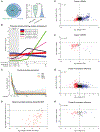
Figure 1.
Figure 1.. The Drosophila Proteome Is Dynamic during Embryogenesis
(A) Venn diagram comparing the previously reported embryonic proteome (Casas-Vila et al., 2017) with that defined in this study. Our methods captured 97% of the genes reported in the previous study and proteins encoded by an additional 2,294 genes. These additional proteins rank significantly lower in abundance than those reported in both studies. A small number of proteins that mapped to multiple genes were excluded from this analysis. (B) k-means clustering (k = 6) for 7,956 quantified proteins through embryogenesis. Embryos were aged to five developmental time points at 22 C. The equivalent developmental times at 25 C for each sample are indicated on the x axis. The first three samples cover the MZT (gray box). (C) Expression profile of individual cluster 6 proteins (gray lines) through embryogenesis reveals the maternal nature of these proteins and their rapid clearance during the MZT, restricting their expression mostly to the early embryo. Profiles of SMG, Cup, TRAL, and ME31B are shown in color, highlighting the later timing of SMG’s degradation relative to its co-repressors. Error bars indicate upper and lower limit of relative expression with a 95% Bayesian confidence interval at each time point. (D) Scatterplot comparing the degradation of cluster 6 proteins during the MZT in this study with that previously reported (Casas-Vila et al., 2017). 104 of our 154 cluster 6 proteins were also found in that study. Almost all cluster 6 proteins that decreased in expression during the MZT (time point 3/time point 1) showed a similar decrease in the previous report (4–5 h/0–1 h) (plotted in red). Note that proteins that decreased to undetectable levels by 4–5 h are plotted on the x axis. (E and F) Scatterplot of change in RNA expression (reads per kilobase of transcript, per million mapped reads [RPKM], 3–4 h/0–1 h) (Eichhorn et al., 2016) versus change in protein expression (time point 3/time point 1) for dynamic proteins during the MZT. Genes corresponding to RNA with R2-fold decrease in expression are plotted in red; R2-fold increase in expression are plotted in blue. (E) Proteins that increased in expression during the MZT (cluster 4) showed a significant correspondence with R2-fold increase in their transcript expression (p < 10 9). (F) Proteins that decreased in expression (cluster 6) showed a significant correspondence with R2-fold decrease in their transcript expression during the MZT (p < 10 22). (G and H) Scatterplot of change in translational efficiency (TE, 3–4 h/0–1 h) (Eichhorn et al., 2016) versus change in protein expression (time point 3/time point 1) for dynamic proteins during the MZT. Genes corresponding to RNA with R2-fold decrease in TE are plotted in red; R2-fold increase in TE is plotted in blue. (G) Cluster 4 proteins showed a significant correspondence with R2-fold increase in TE of their transcripts (p < 10 3). (H) Cluster 6 proteins were not associated with significant changes in TE of their transcripts (p = 0.206).Fisher’s exact test was performed for (E)–(H). See also Figure S1 and Tables S1 and S2.
Figure 2.
Figure 2.. SMG, Cup, TRAL, and ME31B Are Degraded at Distinct Times during the MZT through the Ubiquitin-Proteasome System
(A) Developmental western blot of wild-type embryos collected in 30-min time windows and aged at 15-min intervals over the first 6 h after egg lay (AEL). Cup, TRAL, and ME31B are notably decreased by around 1.5 h AEL, whereas SMG levels increase in the early embryo and are subsequently cleared by about 2.5 h AEL. BEL and eIF4E are present for the duration of the time course. Tubulin was probed as a loading control. See Figure S2 for quantification. (B) Developmental western blot of wild-type embryos collected over the first 4 h of embryogenesis from mated females (fertilized) and unmated females (unfertilized). Degradation of Cup, TRAL, and ME31B are unaffected in unfertilized eggs, whereas SMG protein fails to be degraded by 3–4 h AEL. (C) Western blots of 1- to 2-h-old embryos that were permeabilized and incubated for 3 h in buffer, DMSO control, or 100 mM MG132 and aged to 4–5 h AEL. All four RBPs shown are stabilized by MG132 treatment. See also Figures S2 and S3.
Figure 3.
Figure 3.. SMG Interacts with Repressor RBPs and Two Distinct E3 Ubiquitin Ligase Complexes
(A and B) FLAG IP-MS of 0- to 3-h embryo lysate collected from transgenic flies expressing FLAG-SMG and homozygous for the deletion allele smg47. A combination of Protein A control IP and FLAG IP from non-transgenic embryo lysate was used as control. Average spectral counts are plotted for proteins detected at R1 in FLAG-SMG IP on average across at least four biological replicates. Significance of interactors was analyzed using SAINT and annotated for proteins of interest: *p % 0.1, **p % 0.05, ***p % 0.001, ^Bait (significance of interaction not applicable). (A) In the absence of RNase A, SMG exhibited significant interactions with the co-repressive complex: Cup, TRAL, ME31B, BEL, and eIF4E (red). (B) In the presence of RNase A, SMG retained significant interactions with its co-repressors Cup and TRAL, but not significantly with ME31B (red). The IP also captured RNA-independent interactions with two E3 ubiquitin ligase complexes: the CTLH complex (blue: Muskelin, RanBPM, CG6617, CG3295, CG31357, and CG7611) and the SCF complex (green: CUL1, SKPA, CG14317, and SLMB). (C and D) GFP IP-MS of 0- to 2-h lysate from embryos expressing either Muskelin-GFP or GFP-SLMB. GFP IP from non-transgenic embryo lysate was used as control. Average iBAQ intensities (Cox and Mann, 2008) for proteins across three biological replicates are plotted. Significance of enrichment in IP versus control was analyzed for each interactor using Student’s t test and annotated for proteins of interest: *p % 0.1, **p % 0.05; ***p % 0.01, ^Bait. (C) Muskelin-GFP interacts with the RBPs (red) and other members of the CTLH complex (blue). (D) GFP-SLMB interacts with the RBPs (red) and other members of the SCF complex (green). Proteins not detected in control IPs were assigned an average spectral count of 0.1 (A and B) or an iBAQ value of 10 (C and D) to avoid log(0); these small values were at least 2-fold less than the lowest detected values across all experiments. See also Tables S4 and S5.
Figure 4.
Figure 4.. The CTLH Complex Directs the Degradation of Cup, TRAL, and ME31B, but Not SMG
(A–C) Quantified developmental western blots of RBP expression. Embryos were collected from maternal knockdown of CTLH complex members over the first 4 h AEL. Knockdown of muskelin, ranBPM, and CG3295 each independently resulted in significant stabilization of Cup (A), TRAL (B), and ME31B (C) relative to control mCherry knockdown. (D) SMG protein degradation was unaffected by knockdown of the CTLH complex. *p < 0.05; n = 3; error bars indicate SD, Student’s t test. n.s., not significant. Knockdown was confirmed by qRT-PCR (Figure S3). See also Figures S4 and S5.
Figure 5.
Figure 5.. The SCF Complex Directs the Degradation of SMG, but Not Cup, TRAL, and ME31B
(A–C) Quantified developmental western blots of RBP expression. Embryos were collected from maternal knockdown of SCF complex members over the first 4 h AEL. Cup (A), TRAL (B), and ME31B (C) protein degradation were unaffected by knockdown of the SCF complex. (D) Knockdown of cul1, skpA, and slmb each independently resulted in significant stabilization of SMG protein relative to control mCherry knockdown. *p < 0.05; n = 3; error bars indicate SD, Student’s t test. n.s., not significant. Knockdown was confirmed by qRT-PCR (Figure S6). See also Figure S7.
Figure 6.
Figure 6.. Subunits of the E3 Ligase Com plexes Are Temporally Regulated during the MZT
(A) Expression of subunits of the CTLH complex captured in the developmental proteome. Error bars indicate the upper and lower limit of relative expression with a 95% Bayesian confidence interval at each time point. Most subunits have relatively constant levels throughout embryogenesis, whereas levels of Muskelin are highest at the first time point and then decrease rapidly. (B) Western blot of embryos expressing Muskelin-GFP. Anti-GFP (top) confirmed rapid clearance of Muskelin-GFP from the embryo. Anti-CG3295 (bottom) confirmed its stable expression during the MZT. (C) Expression of subunits of the SCF complex captured in the developmental proteome. Error bars are the same as in (A). Most subunits exhibit relatively constant levels throughout embryogenesis, whereas levels of the F-box subunit CG14317 increased rapidly during the MZT and then decreased very rapidly by the end of the MZT, with peak levels coinciding with degradation of SMG protein. (D) Developmental western blot of control RNAi embryos, confirming the stable expression of SLMB during the MZT. Notably, the same blot shown here was used to confirm SLMB knockdown in Figure S6.
Figure 7.
Figure 7.. Persistent SMG Protein Downre gulates Zygotic Re-expression of Its Target Transcripts
(A) Transgenic flies were generated expressing either FLAG-tagged full-length SMG or SMG767D999 truncated C-terminal to its SAMPHAT RNA-binding domain. Transgenes were under the control of endogenous regulatory elements. (B) Developmental western blots were performed on embryos collected from transgenic flies in the smg47 deletion mutant background. FLAG-SMG expression resembled that of endogenous SMG. SMG767D999 protein was stabilized and persisted through the MZT. (C–E) Embryos were collected from the transgenic flies at two time points during the MZT, and gene expression was assayed by qRT-PCR. (C) Expression of transcripts that depend on SMG for zygotic transcription was rescued by SMG767D999 to similar or higher levels than by full-length SMG. These transcripts are predicted not to be direct targets for SMG binding because they have low SRE scores (see STAR Methods). (D) Degradation of SMG-bound target maternal transcripts was rescued by SMG767D999. Where transcripts were not completely degraded by FLAG-SMG, SMG767D999 persistence resulted in further degradation of these targets to significantly lower levels (rightmost six genes). (E) SMG-target maternal transcripts that are re-expressed zygotically were significantly down-regulated in their zygotic levels by persistence of SMG767D999. Wilcoxon signed rank test p values are shown for each gene group; two biological replicates for each gene; error bars indicate SD.

Similar articles

Cited by

References

    1. Aanes H, Collas P, and Aleström P (2014). Transcriptome dynamics and diversity in the early zebrafish embryo. Brief. Funct. Genomics 13, 95–105. - PubMed
    1. Aviv T, Lin Z, Lau S, Rendl LM, Sicheri F, and Smibert CA (2003). The RNA-binding SAM domain of Smaug defines a new family of post-transcriptional regulators. Nat. Struct. Biol 10, 614–621. - PubMed
    1. Aviv T, Lin Z, Ben-Ari G, Smibert CA, and Sicheri F (2006). Sequence-specific recognition of RNA hairpins by the SAM domain of Vts1p. Nat. Struct. Mol. Biol 13, 168–176. - PubMed
    1. Bailey TL, and Elkan C (1994). Fitting a mixture model by expectation maximization to discover motifs in biopolymers. Proc. Int. Conf. Intell. Syst. Mol. Biol 2, 28–36. - PubMed
    1. Baltz AG, Munschauer M, Schwanhäusser B, Vasile A, Murakawa Y, Schueler M, Youngs N, Penfold-Brown D, Drew K, Milek M, et al. (2012). The mRNA-bound proteome and its global occupancy profile on protein-coding transcripts. Mol. Cell 46, 674–690. - PubMed

Publication types

MeSH terms