. 2020 Jul 6;219(7):e201903031.
doi: 10.1083/jcb.201903031.

A polybasic domain in aPKC mediates Par6-dependent control of membrane targeting and kinase activity

Affiliations

A polybasic domain in aPKC mediates Par6-dependent control of membrane targeting and kinase activity

Wei Dong et al. J Cell Biol. .

Abstract

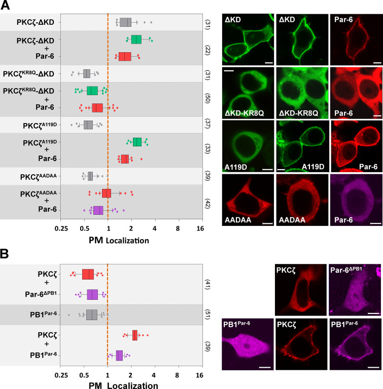
Mechanisms coupling the atypical PKC (aPKC) kinase activity to its subcellular localization are essential for cell polarization. Unlike other members of the PKC family, aPKC has no well-defined plasma membrane (PM) or calcium binding domains, leading to the assumption that its subcellular localization relies exclusively on protein-protein interactions. Here we show that in both Drosophila and mammalian cells, the pseudosubstrate region (PSr) of aPKC acts as a polybasic domain capable of targeting aPKC to the PM via electrostatic binding to PM PI4P and PI(4,5)P2. However, physical interaction between aPKC and Par-6 is required for the PM-targeting of aPKC, likely by allosterically exposing the PSr to bind PM. Binding of Par-6 also inhibits aPKC kinase activity, and such inhibition can be relieved through Par-6 interaction with apical polarity protein Crumbs. Our data suggest a potential mechanism in which allosteric regulation of polybasic PSr by Par-6 couples the control of both aPKC subcellular localization and spatial activation of its kinase activity.


Figures

Figure 1.
Figure 1.
The conserved polybasic PSr mediates PM targeting of aPKC in Drosophila epithelia. (A) Alignment of the PSr (bold) and adjacent sequences in C1 domain from Drosophila and mammalian aPKC isoforms. Sequences are based on NCBI NP_524892.2 (DaPKC), NP_002735.3 (PKCζ), and NP_002731.4 (PKCι). Residues mutated in aPKCKR8Q (KR8Q), aPKCKR8A (KR8A), PKCζA119D (A119D), and PKCζAADAA (AADAA) are also shown. (B) GST fusion of PSr from PKCζ (GST-PSr), but not the nonpolybasic GST-PSr-KR8A, cosedimented with PI4P- and PIP2-containing liposomes in vitro. (C) DaPKC::GFP (DaPKC), but not nonpolybasic DaPKCKR8Q::GFP (KR8Q), localized to PM in embryonic and larval disc epithelia. (D) Follicular epithelial cells in ovaries from ubi-DaPKC::GFP or par-6::GFP adult females were imaged ex vivo under controlled oxygen environment. Cells are in cross-section view as indicated by the illustration at the far right. PM localization of DaPKC::GFP and Par-6::GFP were acutely inhibited by hypoxia (0.5% O2) but recovered after reoxygenation by air (see also Videos 1 and 2). Kymographs on the right show the acute loss and recovery DaPKC::GFP or Par-6::GFP on PM during hypoxia and posthypoxia reoxygenation treatments. White boxes indicate where kymographs were sampled. Scale bars: 5 µm (C and D).
Figure S1.
Figure S1.
Polybasic PSr is required for PM targeting of Drosophila aPKC. (A) PM localization of wild-type DaPKC::GFP and nonpolybasic DaPKCKR8Q::GFP in Drosophila wild type and DaPKC−/− mutant follicular epithelial cells. Asterisks indicate DaPKC−/− mutant cells identified by the loss of Histon2A::RFP (His2A::RFP). Images are in cross-section view. (B) In Drosophila embryonic epithelial cells, PM localization of Par-6::GFP or DaPKC::GFP was lost under hypoxia (0.5% O2) but recovered after posthypoxia reoxygenation. Images are in tangential view of the apical surface of embryonic epithelia. (C) Par-6::GFP showed acute and reversible loss of PM targeting under hypoxia in both wild-type and lgl−/− mutant (marked by the loss of nuclear RFP) follicular epithelial cells. Note that in lgl−/− mutant cells, Par-6 was no longer restricted to apical PM but localized to both apical and lateral PM. Images are in cross-section view. Scale bars: 5 µm.
Figure 2.
Figure 2.
PM localization of PKCζ in HEK293 cells requires both polybasic PSr and Par-6. (A) PKCζ::GFP or Par-6::RFP was cytosolic when expressed alone, but both became strongly PM-localized when coexpressed. PKCζ::RFP-2A and 2A-Par-6::iRFP also showed strong PM localization. (B) Pan-aPKC antibody (anti-aPKC) detects both exogenously expressed PKCL::GFP and PKCζ::GFP in HEK293 cells (white arrowhead), as well as endogenous expressed aPKC (black arrowhead). PKCζ-specific antibody (anti-PKCζ) specifically detected exogenously expressed PKCζ::GFP but showed no detectable expression of endogenous PKCζ in HEK293. α-Tubulin serves as loading control. (C) Only PKCζ::RFP-2A (∼100 kD) but not full-length PKCζ::RFP-2A-Par-6::iRFP fusion protein (∼150 kD) was detected in cells expressing PKCζ::RFP-2A-Par-6::iRFP. Lysate from cells expressing PKCζ::RFP was loaded and blotted as a positive control. (D) PKCζC107Y::GFP (C107Y), PKCζKR8Q::GFP (KR8Q), and PKCζΔPSr::GFP (ΔPSr) did not localize to PM when coexpressed with Par-6::RFP. (E) Both PKCζ::GFP and PKCζKR8A::GFP, but not PKCζC107Y::GFP, coimmunoprecipitated with FLAG-Par-6 by anti-GFP antibody from HEK293 cells. In all data plots, boxes extend from 25th and 75th percentiles, with lines in the middle indicating the median and whiskers indicating 10th and 90th percentiles. Sample numbers are indicated in parentheses at the right. Orange dashed lines in quantification figures indicate that the PM localization index = 1 (see Materials and methods). Measurements <1 indicate cytosolic localization, and those >1 indicate PM localization. PM localization axes in all figures are in log2 scale. Scale bars: 5 µm (A and D).
Figure S2.
Figure S2.
Par-6–dependent PM targeting of PKCζ in MCF7, COS7, and polarized MDCK cells. (A and B) Representative images showing that in MCF7 and COS7 cells, PKCζ and Par-6 were cytosolic when expressed alone, but both became PM-localized when coexpressed. (C) Representative images showing that in polarized MDCK cells, overexpressed PKCζ::GFP was cytosolic and Par-6::RFP was partially PM-localized. Both PKCζ::GFP and Par-6::RFP were PM-localized when coexpressed, whereas both PKCζKR8Q::GFP and Par-6::RFP were cytosolic when coexpressed. Scale bars: 5 µm.
Figure 3.
Figure 3.
PM targeting of PKCζ in HEK293 cells is independent of Cdc42. (A) PKCζ::GFP was cytosolic when coexpressed with BFP::Cdc42CA. Par-6::RFP localized to PM in cells coexpressing GFP::Cdc42CA. Coexpressed PKCζ and Pra-6 were PM-localized in cells expressing either BFP::Cdc42CA or BFP::Cdc42DN. Nonpolybasic PKCζKR8Q::GFP was PM-localized in cells expressing both BFP::Cdc42CA and Par-6::RFP. (B) Overexpression of Cdc42CA did not change DaPKC::GFP and DaPKCKR8Q::GFP subcellular localization in Drosophila follicular epithelial cells. (C) DaPKC::GFP formed intracellular puncta (arrowheads) in cdc42-RNAi follicular cells. In B and C, cells expressing UAS-cdc42-RNAi or UAS-Cdc42CA are marked by RFP expression. Asterisks highlight wild-type cells. Scale bars: 5 µm (A and B); 15 µm (C).
Figure 4.
Figure 4.
PM targeting of PKCζ and Par-6 depends on both PI4P and PIP2. (A) INPP5E converts PIP2 to PI4P, which can be further converted to PI by Sac, whereas PJ converts both PIP2 and PI4P to PI. Box: FKBP-PJ can be acutely recruited to PM through rapamycin (rapa)-induced heterodimerization with PM-anchored Lyn11-FRB. PM recruitment of PJ results in acute depletion of both PI4P and PIP2. (B) PM localization of PKCζ::GFP and Par-6::iRFP was quantified before and after rapamycin addition in HEK293 cells expressing Lyn11-FRB-CFP and mCherry-FKBP-PJ, -Sac, -INPP5E, or -PJ-dead (as a negative control). Representative time-lapse images of Lyn11-FRB-CFP (red), PKCζ::GFP (green), Par-6::iRFP (magenta), and mCherry-FKBP-PJ/Sac/INPP5E/PJ-dead (cyan) are shown under each quantification figure. For each quantification, means ± SEM from 20–30 cells pooled across three independent experiments were plotted. (C) Purified GST-PKCζ/Par-6::His6 complex, but not purified GST-PKCζ alone or GST-PKCζKR8Q/Par-6::His6 complex, bound to PI4P- and PIP2-liposomes. Scale bars: 5 µm (B, all panels).
Figure S3.
Figure S3.
Par-6::GFP expands to basolateral PM in Drosophila follicular epithelial cells under the acute loss of PIP2. Cells overexpressing mRFP-FKBP-INPP5E and PM-bound Lck-FRB::CFP (not depicted) are labeled by nuclear RFP (asterisks in GFP images). Rapamycin (rapa) treatment induced strong PM localization of mRFP-FKBP-INPP5E, but Par-6::GFP remained largely on apical PM (A, cross-section view) with expansion to lateral PM (B, tangential view of basolateral PM). Scale bars: 5 µm.
Figure 5.
Figure 5.
Par-6 interaction with PKCζ is required for polybasic PSr to bind PM. (A) PKCζ-ΔKD::GFP (ΔKD) localized to PM with or without the coexpression of Par-6::RFP. PKCζ-ΔKDKR8Q::GFP (ΔKD-KR8Q) was cytosolic with or without the coexpression of Par-6::RFP. PKCζA119D::GFP (A119D) and PKCζAADAA::GFP (AADAA) were cytosolic when expressed alone. When coexpressing with Par-6::RFP, PKCζA119D::GFP was strongly PM-localized, whereas PKCζAADAA::GFP was barely PM-localized. (B) PKCζ::RFP and Par-6ΔPB1::iRFP remained in cytosol when coexpressed. PB1Par-6::iRFP was cytosolic when expressed alone, but when coexpressed with PKCζ::RFP, both became PM-localized. All experiments were performed in HEK293 cells. Scale bars: 5 µm.
Figure 6.
Figure 6.
PM-targeted PKCζ/Par-6 complex is inhibited from phosphorylating Lgl. (A) PM localization of Lgl::GFP was strongly reduced in cells expressing PKCζ::RFP but not kinase-dead PKCζK281W::RFP. Nonphosphorylatable LglS6A::GFP remained PM-localized in cells expressing PKCζ::RFP. Lgl::GFP remained PM-localized in cells coexpressing PKCζ::RFP and Par-6::iRFP. Lgl::GFP also showed strong PM localization in cells expressing PKCζ::RFP-2A-Par-6::iRFP. (B) PM localization of Lgl::GFP was strongly reduced in cells expressing Lyn11-PKCζ::RFP, PKCζKR8Q::RFP (KR8Q), and Lyn11-PKCζKR8Q::RFP (Lyn11-KR8Q). In all three cases, coexpression of Par-6::iRFP increased PM localization of Lgl::GFP. (C) Cells expressing Lgl::GFP only, expressing both Lgl::GFP and PKCζ::RFP (or PKCζKR8Q::RFP or Lyn11-PKCζKR8Q::RFP), or expressing Lgl::GFP together with PKCζ::RFP (or PKCζKR8Q::RFP or Lyn11-PKCζKR8Q::RFP) and FLAG::Par-6, were directly lysed in SDS loading buffer and analyzed by Western blot. Anti-(P)-Lgl, antibody against phosphorylated Lgl. All experiments were performed in HEK293 cells. Scale bars: 5 µm.
Figure S4.
Figure S4.
Interaction between Par-6 and Crb-intra in HEK293 cells. (A) Par-6::iRFP localized to PM in HEK293 cells expressing BFP::Crb-intra, but not in cells expressing BFP::Crb-intraΔERLI. PKCζKR8Q::RFP was PM-localized in cells expressing Par-6::iRFP and BFP::Crb-intra but was cytosolic in cells expressing Par-6::iRFP and BFP::Crb-intraΔERLI. (B) Lgl::GFP was predominantly PM-localized in polarized MDCK cells overexpressing PKCζ::RFP or PKCζ::RFP-2A-Par-6::iRFP. LglS6A::GFP was PM-localized in MDCK cells expressing PKCζ::RFP–2A-Par-6::iRFP. Scale bars: 5 µm.
Figure 7.
Figure 7.
Crb-intra activates PM-targeted aPKC/Par-6 complex. (A) In HEK293 cells expressing either PB1Par-6::iRFP or Par-6ΔPDZ::iRFP, PKCζ::RFP was strongly PM-localized, whereas Lgl::GFP was strongly reduced from PM. (B) In HEK293 cells expressing Lgl::GFP, PKCζ::RFP, and Par-6::iRFP, PM localization of Lgl::GFP was strongly reduced when BFP::Crb-intra, but not BFP::Crb-intraΔERLI or BFP::Cdc42CA, was coexpressed. LglS6A::GFP remained on PM in cells expressing PKCζ::RFP, Par-6::iRFP, and BFP::Crb-intra. Lgl::GFP remained on PM in cells expressing BFP::Crb-intra, Par-6::iRFP, and kinase-dead PKCζK281W::RFP. Scale bars: 5 µm.
Figure 8.
Figure 8.
Crb promotes DaPKC phosphorylation on Lgl in vivo. (A and B) Drosophila lgl::GFP or lglS6A::GFP follicular epithelial cells overexpressing Crb were immunostained for GFP (green), DaPKC (red), and Crb (magenta). Images in A are in tangential view and were sectioned below the apical surface of follicular cells where Crb and aPKC normally are absent. Images in B are in cross section view of follicular epithelial cells, showing overexpressed Crb expanded into the lateral PM along with DaPKC. Cells overexpressing Crb are highlighted by asterisks in green-channel images. (C) Wild-type lgl::GFP embryos and embryos of lgl::GFP UAS-Crb/Mat-Gal4 or lglS5A::GFP UAS-Crb/Mat-Gal4 were immunostained for GFP (green), Crb (red), and aPKC (magenta). All embryonic epithelial cells were in cross-section view. Note the loss of Lgl::GFP, but not LglS5A::GFP, from the PM under Crb overexpression driven by Mat-Gal4 (see Materials and methods). (D) Wild-type lgl::GFP embryos and lgl::GFP; crb−/− mutant embryos were immunostained for GFP (green), Crb (red), and aPKC (magenta). In crb−/− embryos, red channel was overexposed to confirm no detectable expression of Crb. In crb−/− embryonic epithelial cells, both Lgl and DaPKC became localized all around PM. (E and F) Lgl::GFP remained on PM in Crb-overexpressing cells that also expressed DaPKC-RNAi (E) or par-6-RNAi (F). (G) Lgl::GFP was severely lost from PM in follicular cells expressing DaPKCΔN. In E–G, cells expressing DaPKC-RNAi, par-6-RNAi or DaPKCΔN are marked by RFP expression (see Materials and methods). Scale bars: 5 µm.
Figure 9.
Figure 9.
A hypothetic model of aPKC PM targeting and kinase activation. (A) Free cytosolic aPKC in autoinhibited conformation has polybasic PSr blocked by the KD from binding to PM. Binding of Par-6 to aPKC induces conformation changes that expose the PSr in aPKC and allow the C-terminus of Par-6 to simultaneously inhibit the aPKC KD. (B) Polybasic PSr in aPKC/Par-6 complex binds to PM via electrostatic interaction with PI4P and PIP2, which are uniquely enriched on PM. (C) Intracellular domain of apical polarity protein Crb interacts with the C-terminal PDZ domain of Par-6 and releases its inhibition on aPKC KD. Interaction with Crb could also facilitate the apical enrichment of PM-bound aPKC/Par-6 in cells. (D) Activated aPKC phosphorylates Lgl to prevent it from binding to apical PM. Illustration is based on Drosophila epithelial cells. AJ, adherens junction.

Similar articles

Cited by

References

    1. Arbuzova A., Schmitz A.A., and Vergères G.. 2002. Cross-talk unfolded: MARCKS proteins. Biochem. J. 362:1–12. 10.1042/bj3620001 - DOI - PMC - PubMed
    1. Atwood S.X., Chabu C., Penkert R.R., Doe C.Q., and Prehoda K.E.. 2007. Cdc42 acts downstream of Bazooka to regulate neuroblast polarity through Par-6 aPKC. J. Cell Sci. 120:3200–3206. 10.1242/jcs.014902 - DOI - PMC - PubMed
    1. Bailey M.J., and Prehoda K.E.. 2015. Establishment of Par-Polarized Cortical Domains via Phosphoregulated Membrane Motifs. Dev. Cell. 35:199–210. 10.1016/j.devcel.2015.09.016 - DOI - PMC - PubMed
    1. Betschinger J., Mechtler K., and Knoblich J.A.. 2003. The Par complex directs asymmetric cell division by phosphorylating the cytoskeletal protein Lgl. Nature. 422:326–330. 10.1038/nature01486 - DOI - PubMed
    1. Brzeska H., Guag J., Remmert K., Chacko S., and Korn E.D.. 2010. An experimentally based computer search identifies unstructured membrane-binding sites in proteins: application to class I myosins, PAKS, and CARMIL. J. Biol. Chem. 285:5738–5747. 10.1074/jbc.M109.066910 - DOI - PMC - PubMed

Publication types

MeSH terms