Review
. 2020 Jun 22;10(6):937.
doi: 10.3390/biom10060937.

Exploring the Role of Non-Coding RNAs in the Pathophysiology of Systemic Lupus Erythematosus

Affiliations
Review

Exploring the Role of Non-Coding RNAs in the Pathophysiology of Systemic Lupus Erythematosus

Mohammad Taheri et al. Biomolecules. .

Abstract

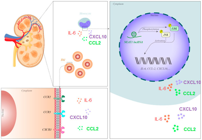
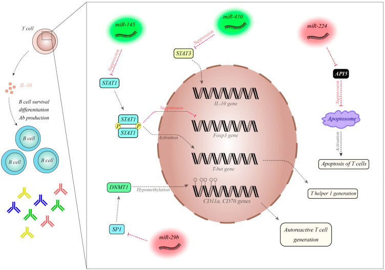
Systemic lupus erythematosus (SLE) is a chronic immune-related disorder designated by a lack of tolerance to self-antigens and the over-secretion of autoantibodies against several cellular compartments. Although the exact pathophysiology of SLE has not been clarified yet, this disorder has a strong genetic component based on the results of familial aggregation and twin studies. Variation in the expression of non-coding RNAs has been shown to influence both susceptibility to SLE and the clinical course of this disorder. Several long non-coding RNAs (lncRNAs) such as GAS5, MALAT1 and NEAT1 are dysregulated in SLE patients. Moreover, genetic variants within lncRNAs such as SLEAR and linc00513 have been associated with risk of this disorder. The dysregulation of a number of lncRNAs in the peripheral blood of SLE patients has potentiated them as biomarkers for diagnosis, disease activity and therapeutic response. MicroRNAs (miRNAs) have also been shown to affect apoptosis and the function of immune cells. Taken together, there is a compelling rationale for the better understanding of the involvement of these two classes of non-coding RNAs in the pathogenesis of SLE. Clarification of the function of these transcripts has the potential to elucidate the molecular pathophysiology of SLE and provide new opportunities for the development of targeted therapies for this disorder.

Keywords: lncRNA; miRNA; systemic lupus erythematosus.


Conflict of interest statement

The authors declare they have no conflict of interest.

Figures

Figure 1
Figure 1
NEAT1 has been shown to be over-expressed in the monocytes of SLE patients. This lncRNA phosphorylates JNK and ERK, thus activating these proteins and enhancing the expression of IL-6, CCL2 and CXCL10. These cytokines/chemokines attract Th1 cells, thus participating in the pathogenesis of nephritis [22]. Abbreviations: SLE, systemic lupus erythematosus; lncRNA, long non-coding RNA, Il-6: interleukin-6, CCL2: C-C motif ligand 2, CXCL10: C-X-C motif chemokine 10.
Figure 2
Figure 2
MiR-145 is decreased in SLE patients. MiR-145 has a role in the inhibition of STAT1, which is involved in the differentiation of Treg cells via its inhibition of Foxp3. Moreover, STAT1 induces T-bet, which participates in the differentiation of Th1 cells [36]. MiR-410 is downregulated in SLE patients and is a suppressor of STAT3. STAT3 is an inducer of IL-10. The levels of this cytokine are increased in SLE patients, leading to the enhanced survival and differentiation of B cells and the increased production of antibodies [37]. MiR-224 is increased in SLE patients and has been shown to inhibit API5. API5 decreases the expression of APAF1 and precludes the activation of caspases, thereby hampering apoptosome formation. Thus, the over-expression of miR-224 leads to increased T cell activation-induced cell death [36]. MiR-29a is upregulated in SLE patients and suppresses Sp1, which is an inducer of ANMT1. The over-expression of miR-29a leads to hypomethylation of CD11a and CD70, which are involved in the production of autoreactive T cells [38] Abbreviations: Treg cell: Regulatory T cell, DNMT1: DNA (cytosine-5)-methyltransferase 1, SP1: specificity protein 1.

Similar articles

Cited by

References

    1. Ramos P.S., Brown E.E., Kimberly R., Langefeld C.D. Genetic Factors Predisposing to Systemic Lupus Erythematosus and Lupus Nephritis. Semin. Nephrol. 2010;30:164–176. doi: 10.1016/j.semnephrol.2010.01.007. - DOI - PMC - PubMed
    1. Block S.R. A brief history of twins. Lupus. 2006;15:61–64. doi: 10.1191/0961203306lu2263ed. - DOI - PubMed
    1. Deafen D., Escalante A., Weinrib L., Horwitz D., Bachman B., Roy-Burman P., Walker A., Mack T.M. A revised estimate of twin concordance in systemic lupus erythematosus. ARHEAW. 1992;35:311–318. doi: 10.1002/art.1780350310. - DOI - PubMed
    1. Grumet F.C., Coukell A., Bodmer J.G., Bodmer W., McDevitt H.O. Histocompatibility (HL-A) Antigens Associated with Systemic Lupus Erythematosus. New Engl. J. Med. 1971;285:193–196. doi: 10.1056/NEJM197107222850403. - DOI - PubMed
    1. Brown E.E., Edberg J.C., Kimberly R. Fc receptor genes and the systemic lupus erythematosus diathesis. J. Autoimmun. 2007;40:567–581. doi: 10.1080/08916930701763710. - DOI - PubMed

MeSH terms