Review
. 2020 Jun 22;9(6):1514.
doi: 10.3390/cells9061514.

Huntington's Disease-An Outlook on the Interplay of the HTT Protein, Microtubules and Actin Cytoskeletal Components

Affiliations
Review

Huntington's Disease-An Outlook on the Interplay of the HTT Protein, Microtubules and Actin Cytoskeletal Components

Aleksandra S Taran et al. Cells. .

Abstract

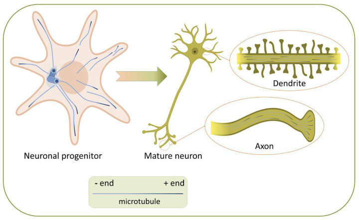
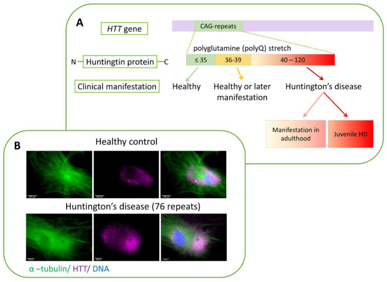
Huntington's disease is a severe and currently incurable neurodegenerative disease. An autosomal dominant mutation in the Huntingtin gene (HTT) causes an increase in the polyglutamine fragment length at the protein N-terminus. The consequence of the mutation is the death of neurons, mostly striatal neurons, leading to the occurrence of a complex of motor, cognitive and emotional-volitional personality sphere disorders in carriers. Despite intensive studies, the functions of both mutant and wild-type huntingtin remain poorly understood. Surprisingly, there is the selective effect of the mutant form of HTT even on nervous tissue, whereas the protein is expressed ubiquitously. Huntingtin plays a role in cell physiology and affects cell transport, endocytosis, protein degradation and other cellular and molecular processes. Our experimental data mining let us conclude that a significant part of the Huntingtin-involved cellular processes is mediated by microtubules and other cytoskeletal cell structures. The review attempts to look at unresolved issues in the study of the huntingtin and its mutant form, including their functions affecting microtubules and other components of the cell cytoskeleton.

Keywords: Huntington’s disease; cytoskeleton; microtubules; neurodegenerative diseases; proteinopathies.


Conflict of interest statement

The authors declare no competing interests.

Figures

Figure 1
Figure 1
Microtubule organization in neuronal progenitor and mature neuron. In the early stages of neuron polarization, MTs originate from the centrosome, where their minus-ends are located during nucleation and elongation before detachment from the centrosome (or oriented after detachment). Hence, the structure of MTs in neuronal progenitor is radial. When neurites mature into axons and dendrites, the structure of the MT network changes. In dendrites, MTs became antiparallel: unique feature of the dendritic MT cytoskeleton is the appearance of MTs with their minus-ends oriented towards the dendritic tips. Simultaneously, in the axons, all the minus-ends are directed towards the body of the neuron.
Figure 2
Figure 2
(A) The correlation of the number of CAG repeats in the HTT gene with the clinical manifestation of HD. The number of repeats ≤35 is normal; from 36 to 39 may not phenotypically manifest (or lead to the development of HD at a later age); from 40 upwards in 100% of cases causes HD. Hence, the age of disease onset and the severity of clinical symptoms depend on the number of CAG repeats according to the inverse correlation principle. (B) Immunocytochemical detection of HTT in the skin fibroblasts of healthy donors and patients with HD. Localization of HTT in the cells of a healthy donor (top panel) and a patient with HD (bottom panel).
Figure 3
Figure 3
Participation of HTT in microtubule transport. HTT functions as a scaffold for the dynein-dynactin complex. (A) Vesicular transport. The phosphorylation of HTT determines the movement direction of the vesicles to the plus or minus end of the MT. (B) The formation of the primary cilia. HTT as a part of the dynein dynactin/HAP1 complex transports PCM1 protein to the basal body, which is necessary for the formation and lengthening of the primary cilia. Anterograde and retrograde transport using molecular motors inside the cilia allows the regulation of the number of receptors on its surface, for example, ARL13B, a small G protein from the Ras superfamily specific for the primary cilia. (C) Mitotic spindle orientation is adjusted using HTT. During cell division HTT is directed to the spindle poles that determines the accumulation of NUMA and LGN there. In addition, HTT controls NUMA and LGN transport along astral microtubules to the cell cortex. At the cell cortex this complex provides pulling forces on astral microtubules for mitotic spindle orientation. HAP1—huntingtin-associated protein 1; PCM1—pericentriolar material 1 protein; ARL13B—ADP-ribosylation factor-like protein 13B; LGN (GPSM2)—leucine-glycine-asparagine (G-protein-signaling modulator 2); NUMA—nuclear mitotic apparatus protein.

Similar articles

Cited by

References

    1. Penazzi L., Bakota L., Brandt R. Microtubule dynamics in neuronal development, plasticity, and neurodegeneration. Int. Rev. Cell Mol. Biol. 2016;321:89–169. doi: 10.1016/bs.ircmb.2015.09.004. - DOI - PubMed
    1. Meiring J.C., Akhmanova A. Microtubules keep large cells in shape. J. Cell Biol. 2020;219:202004031. doi: 10.1083/jcb.202004031. - DOI - PMC - PubMed
    1. Alieva I.B. Role of microtubule cytoskeleton in regulation of endothelial barrier function. Biochemistry. 2014;79:964–975. doi: 10.1134/S0006297914090119. - DOI - PubMed
    1. Dugina V., Alieva I., Khromova N., Kireev I.I., Gunning P.W., Kopnin P.B. Interaction of microtubules with the actin cytoskeleton via cross-talk of EB1-containing +TIPs and γ-actin in epithelial cells. Oncotarget. 2016;7:72699–72715. doi: 10.18632/oncotarget.12236. - DOI - PMC - PubMed
    1. Kopf A., Renkawitz J., Hauschild R., Girkontaite I., Tedford K., Merrin J., Thorn-Seshold O., Trauner D., Häcker H., Fischer K.-D., et al. Microtubules control cellular shape and coherence in amoeboid migrating cells. J. Cell Biol. 2020;219:219. doi: 10.1083/jcb.201907154. - DOI - PMC - PubMed

Publication types