Review
. 2020 Jun 22;12(6):1397.
doi: 10.3390/polym12061397.

Stimuli-Responsive Polymeric Nanocarriers for Drug Delivery, Imaging, and Theragnosis

Affiliations
Review

Stimuli-Responsive Polymeric Nanocarriers for Drug Delivery, Imaging, and Theragnosis

Sabya Sachi Das et al. Polymers (Basel). .

Abstract

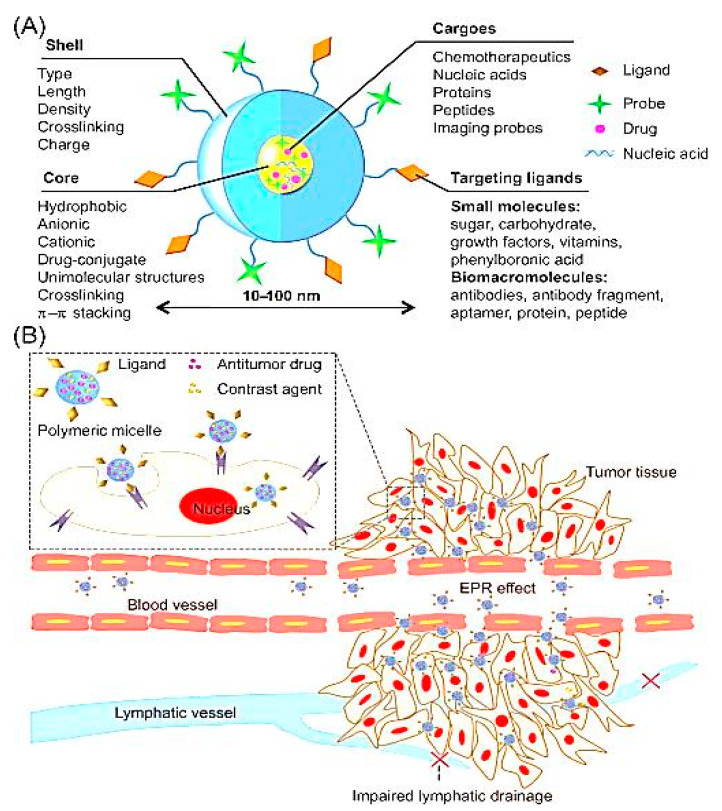
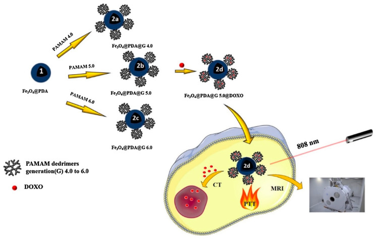
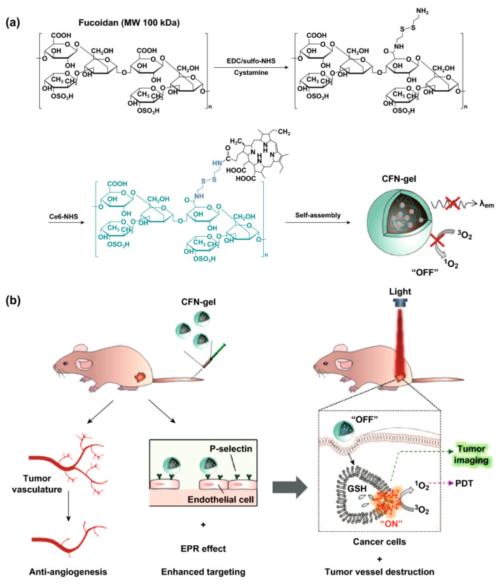
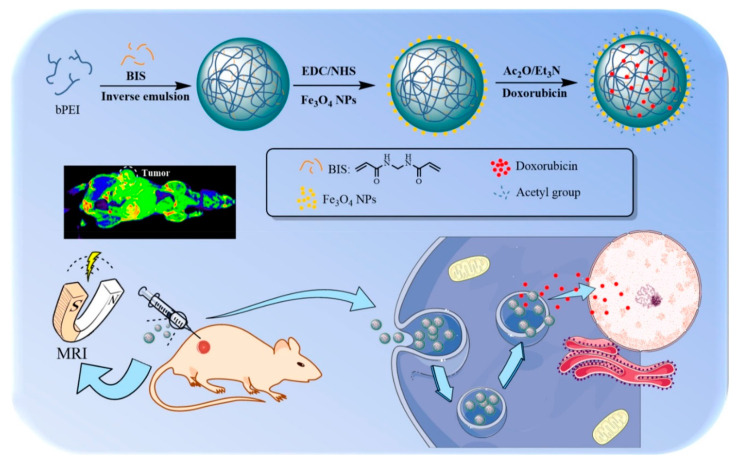
In the past few decades, polymeric nanocarriers have been recognized as promising tools and have gained attention from researchers for their potential to efficiently deliver bioactive compounds, including drugs, proteins, genes, nucleic acids, etc., in pharmaceutical and biomedical applications. Remarkably, these polymeric nanocarriers could be further modified as stimuli-responsive systems based on the mechanism of triggered release, i.e., response to a specific stimulus, either endogenous (pH, enzymes, temperature, redox values, hypoxia, glucose levels) or exogenous (light, magnetism, ultrasound, electrical pulses) for the effective biodistribution and controlled release of drugs or genes at specific sites. Various nanoparticles (NPs) have been functionalized and used as templates for imaging systems in the form of metallic NPs, dendrimers, polymeric NPs, quantum dots, and liposomes. The use of polymeric nanocarriers for imaging and to deliver active compounds has attracted considerable interest in various cancer therapy fields. So-called smart nanopolymer systems are built to respond to certain stimuli such as temperature, pH, light intensity and wavelength, and electrical, magnetic and ultrasonic fields. Many imaging techniques have been explored including optical imaging, magnetic resonance imaging (MRI), nuclear imaging, ultrasound, photoacoustic imaging (PAI), single photon emission computed tomography (SPECT), and positron emission tomography (PET). This review reports on the most recent developments in imaging methods by analyzing examples of smart nanopolymers that can be imaged using one or more imaging techniques. Unique features, including nontoxicity, water solubility, biocompatibility, and the presence of multiple functional groups, designate polymeric nanocues as attractive nanomedicine candidates. In this context, we summarize various classes of multifunctional, polymeric, nano-sized formulations such as liposomes, micelles, nanogels, and dendrimers.

Keywords: drug delivery; endogenous stimuli; exogenous stimuli; imaging; stimuli-responsive; stimuli-responsive targeting; theranostic.


Conflict of interest statement

The authors declare no conflict of interest.

Figures

Figure 1
Figure 1
Depiction of the synthetic stages for the synthesis of UCNPs, their water exchange and two PSs functionalization (Rose Bengal and Chlorin e6). Reproduced from [232] with permission from Springer Nature, 2020.
Figure 2
Figure 2
(A) Illustration depicting the formation of NB-PTXLp conjugates by EDC/NHS coupling, (B) Transmission electron micrograph of NB-PTXLp conjugate (scale bar—200 nm), (C) Transmission electron micrograph of NB/PTXLp conjugate (scale bar—100 nm). Reproduced from [234] with permission from American Chemical Society, 2019.
Figure 3
Figure 3
Design and synthesis of fluorinated probe. (a) Structure of polymer and nanogel. (i) Nanogel formation via crosslinking of PDS groups with DTT (ii) Cleavage of THP group in the presence of HCl and formation of negatively charged moiety with NaOH addition. (b) Schematic representation of preparation of fluorinated nanogel with decreased interior density. Reproduced from [242] with permission from American Chemical Society, 2019.
Figure 4
Figure 4
Polymeric micelles for cancer theranostics. (A) The composition of polymeric micelles. (B) Polymeric micelles target tumor tissues for cancer theranostics, and can be specifically accumulated in tumor tissues through the EPR effect for cancer diagnosis and therapy. EPR: Enhanced permeability and retention. Reproduced from [273] with permission from Elsevier, 2019.
Figure 5
Figure 5
The schematic synthesis presentation of theranostic nanoplatform based on the Fe3O4@PDA@G 4.0–6.0 nanoparticles and its application in combined CT-PTT therapy on HepG2 cells. Reproduced from [300] with permission from Elsevier, 2019.
Figure 6
Figure 6
(a) Synthetic steps of Ce6–fucoidan theranostic nanogel (CFN-gel), (b) schematic illustration of CFN-gel and its mode of action. Reproduced from [305] with permission from Springer Nature, 2020.
Figure 7
Figure 7
Schematic representation of the synthesis of Fe3O4/PEI-Ac NGs/DOX complexes for MR imaging-guided chemotherapy of tumors. Reproduced from [306] with permission from American Chemical Society, 2020.

References

    1. Kundu J.K., Surh Y.-J. Nrf2-Keap1 Signaling as a Potential Target for Chemoprevention of Inflammation-Associated Carcinogenesis. Pharm. Res. 2010;27:999–1013. doi: 10.1007/s11095-010-0096-8. - DOI - PubMed
    1. Mura S., Nicolas J., Couvreur P. Stimuli-responsive nanocarriers for drug delivery. Nat. Mater. 2013;12:991–1003. doi: 10.1038/nmat3776. - DOI - PubMed
    1. Lee E.S., Oh K.T., Kim D., Youn Y.S., Bae Y.H. Tumor pH-responsive flower-like micelles of poly(L-lactic acid)-b-poly(ethylene glycol)-b-poly(L-histidine) J. Control. Release. 2007;123:19–26. doi: 10.1016/j.jconrel.2007.08.006. - DOI - PMC - PubMed
    1. Neri D., Supuran C.T. Interfering with pH regulation in tumours as a therapeutic strategy. Nat. Rev. Drug Discov. 2011;10:767–777. doi: 10.1038/nrd3554. - DOI - PubMed
    1. Chawla J.S., Amiji M.M. Biodegradable poly(ε-caprolactone) nanoparticles for tumor-targeted delivery of tamoxifen. Int. J. Pharm. 2002;249:127–138. doi: 10.1016/S0378-5173(02)00483-0. - DOI - PubMed

LinkOut - more resources