Review
. 2020 Jun 22;21(12):4440.
doi: 10.3390/ijms21124440.

Progressive Skeletal Muscle Atrophy in Muscular Dystrophies: A Role for Toll-like Receptor-Signaling in Disease Pathogenesis

Affiliations
Review

Progressive Skeletal Muscle Atrophy in Muscular Dystrophies: A Role for Toll-like Receptor-Signaling in Disease Pathogenesis

Boel De Paepe. Int J Mol Sci. .

Abstract

Muscle atrophy is an active process controlled by specific transcriptional programs, in which muscle mass is lost by increased protein degradation and/or decreased protein synthesis. This review explores the involvement of Toll-like receptors (TLRs) in the muscle atrophy as it is observed in muscular dystrophies, disorders characterized by successive bouts of muscle fiber degeneration and regeneration in an attempt to repair contraction-induced damage. TLRs are defense receptors that detect infection and recognize self-molecules released from damaged cells. In muscular dystrophies, these receptors become over-active, and are firmly involved in the sustained chronic inflammation exhibited by the muscle tissue, via their induction of pro-inflammatory cytokine expression. Taming the exaggerated activation of TLR2/4 and TLR7/8/9, and their downstream effectors in particular, comes forward as a therapeutic strategy with potential to slow down disease progression.

Keywords: Toll-like receptors; muscular dystrophies; myositis.


Conflict of interest statement

The author declares no conflict of interest. The funders had no role in the design of the study; in the collection, analyses, or interpretation of data; in the writing of the manuscript, or in the decision to publish the results.

Figures
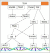
Figure 1
Figure 1
Signaling routes downstream of TLR4. Abbreviations: Activator protein-1 (AP-1), inhibitor of κB kinase (IKK), interleukin-1 receptor-associated kinase (IRAK), interferon regulatory factor (IRF), mitogen activated protein kinase (MAPK), myeloid differentiation primary response 88 (MyD88), nuclear factor κB (NFκB), receptor-interacting protein (RIP), Transforming growth factor-β-activating kinase (TAK), tumor necrosis factor receptor-associated factor (TANK), Toll/IL-1 receptor domain-containing adaptor protein (TIRAP), Toll-like receptor (TLR), tumor necrosis factor receptor-associated factor (TRAF), Toll/IL-1 receptor domain-containing adapter protein inducing interferon-β-related adapter molecule (TRAM), Toll/IL-1 receptor domain-containing adapter protein inducing interferon-β (TRIF).
Figure 2
Figure 2
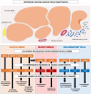
Model of TLR/ligand-driven tissue damage in muscular dystrophies. Proposed mechanism of perpetuated muscle damage in muscular dystrophies: Muscle contractions induce sustained muscle fiber injury that becomes chronic and can no longer be repaired. Due to unsealing of the muscle fiber membranes, intracellular acute-phase protein serum amyloid A1 (SAA1), heat shock protein 70 (HSP70), and high mobility group Box 1 (HMGB1) are released, and become Toll-like receptor (TLR) pathway-activating danger-associated molecular patterns (DAMPs). The corresponding cell surface receptors TLR2 and TLR4 are overexpressed in dystrophic muscle fibers, blood vessels, and tissue-infiltrating inflammatory cells which, upon stimulation, activate proteolytic and inflammatory processes, amplifying tissue damage further. Necrotic muscle fibers release nucleic acids that activate the corresponding cytoplasmic receptors. Released single-stranded RNA (ssRNA) sequences are picked up by blood vessels and inflammatory cells and bind to their receptor TLR7, activating degeneration/regeneration-focused immune cell function. Extracellular mitochondrial DNA (mtDNA) is engulfed by muscle fibers and inflammatory cells that possess its receptor TLR9, which subsequently leads to activation of autophagic processes and stimulates inflammatory responses even further.

References

    1. Emery A.E. The muscular dystrophies. Lancet. 2002;359:687–695. doi: 10.1016/S0140-6736(02)07815-7. - DOI - PubMed
    1. Mosquiera M., Zeiger U., Foerderer M., Brinkmeier H., Fink R.H.A. Cardiac and respiratory dysfunction in Duchenne muscular dystrophy and the role of second messengers. Med. Res. Rev. 2013;5:1174–1213. doi: 10.1002/med.21279. - DOI - PubMed
    1. Mercuri E., Boennemann C.G., Muntoni F. Muscular dystrophies. Lancet. 2019;394:2025–2038. doi: 10.1016/S0140-6736(19)32910-1. - DOI - PubMed
    1. Mouisel E., Vignaud A., Hourde C., Butler-Browne G., Ferry A. Muscle weakness and atrophy are associated with decreased regenerative capacity and changes in MTOR signaling in skeletal muscle of vednerable (18-24-month-old) dystrophic mdx mice. Muscle Nerve. 2010;41:809–818. doi: 10.1002/mus.21624. - DOI - PubMed
    1. Madaro L., Bouche M. From innate to adaptive immune response in muscular dystrophies and skeletal muscle regeneration: The role of lymphocytes. BioMed Res. Int. 2014;2014:438675. doi: 10.1155/2014/438675. - DOI - PMC - PubMed

Substances