

Mueller Matrix Analysis of Collagen and Gelatin Containing Samples Towards More Objective Skin Tissue Diagnostics
- PMID: 32580462
- PMCID: PMC7361993
- DOI: 10.3390/polym12061400
Mueller Matrix Analysis of Collagen and Gelatin Containing Samples Towards More Objective Skin Tissue Diagnostics
Abstract
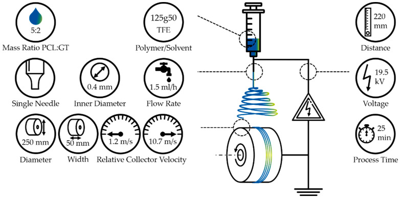
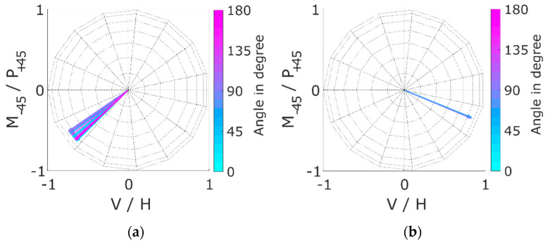
Electrospun polycaprolactone:gelatin (PCL:GT) fibre scaffolds are widely employed in the field of tissue implants. Here, the orientation of fibres plays an important role in regard to implantation due to the impact on the mechanical properties. Likewise, the orientation of collagen fibres in skin tissue is relevant for dermatology. State-of-the-art fibre orientation measurement methods like electron microscopy are time consuming and destructive. In this work, we demonstrate polarimetry as a non-invasive approach and evaluate its potential by measuring the Mueller matrix (MM) of gelatin and collagen containing samples as simple skin tissue phantoms. We demonstrate that it is possible to determine the orientation of PCL:GT fibre scaffolds within one MM measurement. Furthermore, we determine the structural orientation in collagen film samples. Currently, the diagnosis of skin diseases is often performed by image analysis or histopathology respectively, which are either subjective or invasive. The method presented, here, provides an interesting alternative approach for such investigations. Our findings indicate that the orientation of collagen fibres within skin lesions might be detectable by MM measurements in the future, which is of interest for skin diagnostics, and will be further investigated during the next step.
Keywords: Mueller matrix; Raman spectroscopy; collagen; electrospinning; fibre alignment; gelatin; tissue engineering.
Conflict of interest statement
The authors declare no conflicts of interest.
Figures


Similar articles
-
Mueller Matrix Measurement of Electrospun Fiber Scaffolds for Tissue Engineering.Polymers (Basel). 2019 Dec 11;11(12):2062. doi: 10.3390/polym11122062. Polymers (Basel). 2019. PMID: 31835798 Free PMC article.
-
Evaluation of nanofibrous scaffolds obtained from blends of chitosan, gelatin and polycaprolactone for skin tissue engineering.Int J Biol Macromol. 2017 Sep;102:1174-1185. doi: 10.1016/j.ijbiomac.2017.05.004. Epub 2017 May 6. Int J Biol Macromol. 2017. PMID: 28487195
-
Surface modification of nanofibrous polycaprolactone/gelatin composite scaffold by collagen type I grafting for skin tissue engineering.Mater Sci Eng C Mater Biol Appl. 2014 Jan 1;34:402-9. doi: 10.1016/j.msec.2013.09.043. Epub 2013 Oct 5. Mater Sci Eng C Mater Biol Appl. 2014. PMID: 24268275
-
Electrospun gelatin/PCL and collagen/PCL scaffolds for modulating responses of bone marrow endothelial progenitor cells.Exp Ther Med. 2019 May;17(5):3717-3726. doi: 10.3892/etm.2019.7387. Epub 2019 Mar 13. Exp Ther Med. 2019. PMID: 30988757 Free PMC article.
-
Mueller matrix polarimetry for characterization of skin tissue samples: A review.Photodiagnosis Photodyn Ther. 2020 Jun;30:101708. doi: 10.1016/j.pdpdt.2020.101708. Epub 2020 Mar 4. Photodiagnosis Photodyn Ther. 2020. PMID: 32145374 Review.
Cited by
-
Impact of Apparatus Orientation and Gravity in Electrospinning-A Review of Empirical Evidence.Polymers (Basel). 2020 Oct 22;12(11):2448. doi: 10.3390/polym12112448. Polymers (Basel). 2020. PMID: 33105879 Free PMC article. Review.
-
Registration of polarimetric images for in vivo skin diagnostics.J Biomed Opt. 2022 Aug;27(9):096001. doi: 10.1117/1.JBO.27.9.096001. J Biomed Opt. 2022. PMID: 36042549 Free PMC article.
-
Label-free distinction of implant infection-associated bacterial biofilms by Mueller matrix polarimetry.J Biomed Opt. 2025 Aug;30(8):085001. doi: 10.1117/1.JBO.30.8.085001. Epub 2025 Aug 22. J Biomed Opt. 2025. PMID: 40861500 Free PMC article.
-
Computational Modeling of Chromatin Fiber to Characterize Its Organization Using Angle-Resolved Scattering of Circularly Polarized Light.Polymers (Basel). 2021 Oct 5;13(19):3422. doi: 10.3390/polym13193422. Polymers (Basel). 2021. PMID: 34641237 Free PMC article.
-
In Situ Characterization of Polycaprolactone Fiber Response to Quasi-Static Tensile Loading in Scanning Electron Microscopy.Polymers (Basel). 2021 Jun 24;13(13):2090. doi: 10.3390/polym13132090. Polymers (Basel). 2021. PMID: 34202874 Free PMC article.
References
-
- Meinhardt-Wollweber M., Heratizadeh A., Basu C., Günther A., Schlangen S., Werfel T., Schacht V., Emmert S., Haenssle H.A., Roth B. A non-contact remote digital dermoscope to support cancer screening and diagnosis of inflammatory skin disease. Biomed. Phys. Eng. Express. 2017;3:55005. doi: 10.1088/2057-1976/aa84d3. - DOI
-
- Fricke D., Denker E., Heratizadeh A., Werfel T., Wollweber M., Roth B. Non-Contact Dermatoscope with Ultra-Bright Light Source and Liquid Lens-Based Autofocus Function. Appl. Sci. 2019;9:2177. doi: 10.3390/app9112177. - DOI
LinkOut - more resources
Full Text Sources
Miscellaneous
