. 2020 Jun 24;21(1):152.
doi: 10.1186/s13059-020-02064-6.

Single-cell transcriptome and antigen-immunoglobin analysis reveals the diversity of B cells in non-small cell lung cancer

Affiliations

Single-cell transcriptome and antigen-immunoglobin analysis reveals the diversity of B cells in non-small cell lung cancer

Jian Chen et al. Genome Biol. .

Abstract

Background: Malignant transformation and progression of cancer are driven by the co-evolution of cancer cells and their dysregulated tumor microenvironment (TME). Recent studies on immunotherapy demonstrate the efficacy in reverting the anti-tumoral function of T cells, highlighting the therapeutic potential in targeting certain cell types in TME. However, the functions of other immune cell types remain largely unexplored.

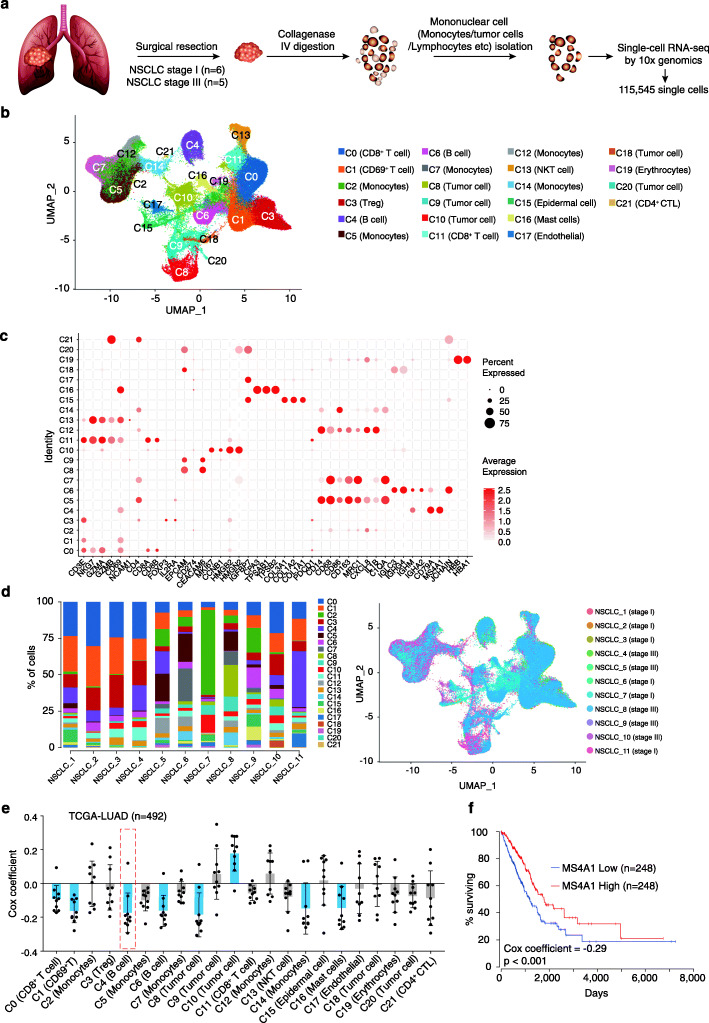
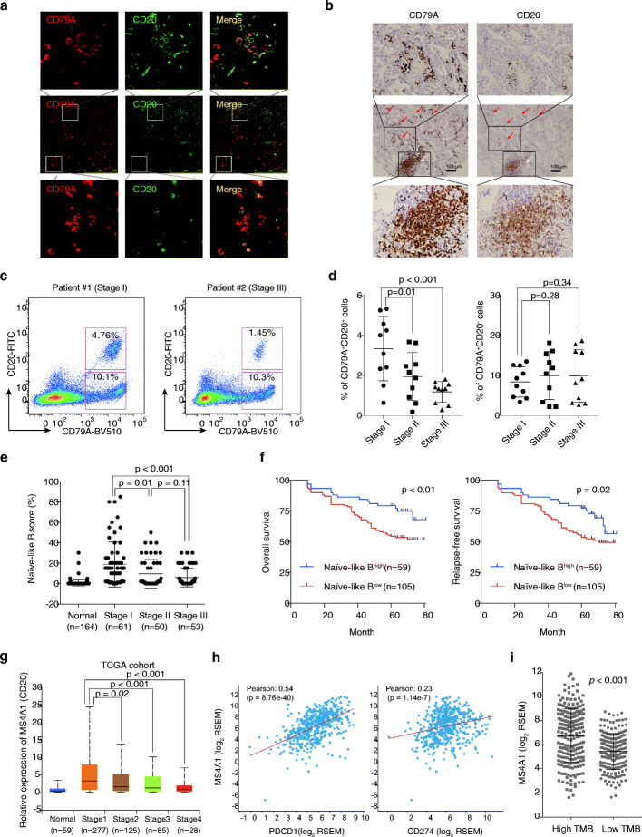
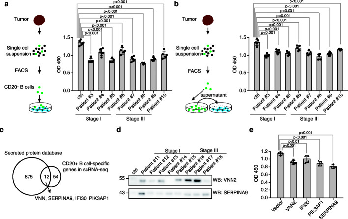
Results: We conduct a single-cell RNA-seq analysis of cells isolated from tumor tissue samples of non-small cell lung cancer (NSCLC) patients, and identify subtypes of tumor-infiltrated B cells and their diverse functions in the progression of NSCLC. Flow cytometry and immunohistochemistry experiments on two independent cohorts confirm the co-existence of the two major subtypes of B cells, namely the naïve-like and plasma-like B cells. The naïve-like B cells are decreased in advanced NSCLC, and their lower level is associated with poor prognosis. Co-culture of isolated naïve-like B cells from NSCLC patients with two lung cancer cell lines demonstrate that the naïve-like B cells suppress the growth of lung cancer cells by secreting four factors negatively regulating the cell growth. We also demonstrate that the plasma-like B cells inhibit cancer cell growth in the early stage of NSCLC, but promote cell growth in the advanced stage of NSCLC. The roles of the plasma-like B cell produced immunoglobulins, and their interacting proteins in the progression of NSCLC are further validated by proteomics data.

Conclusion: Our analysis reveals versatile functions of tumor-infiltrating B cells and their potential clinical implications in NSCLC.


Conflict of interest statement

The authors declare no competing interests.

Figures

Fig. 1
Fig. 1
Single-cell transcriptomic analysis reveals the transcriptome of cells in the microenvironment of lung cancer. a Schematic illustration of the experimental design of this study. b Visualization of single-cell RNA-seq data of 115,545 cells freshly isolated primary lung cancer by UMAP. c Representative gene expression in 22 subclasses of cells. d Distribution of the 22 subclasses among 11 patients with NSCLC. e, f The prognosis value of the genes uniquely expressed in each class of the 22 cell types
Fig. 2
Fig. 2
Co-existence of the two major subtypes of B cells in the microenvironment of NSCLC tumors. a Schematic illustration of the analysis procedure. b Visualization of single-cell RNA-seq data of B cells by UMAP. c Representative gene expression among different classes of B cells. d Expression of MS4A1 (CD20), CD22, CD19, TNFRSF17 (BCMA), IGHG1, and IGHG4 in B cells. e Distribution of different subclasses of B cells in stage I and stage III NSCLC. f Heatmap showing the difference of BC1 (naïve-like B cells) between NSCLC and PBMC
Fig. 3
Fig. 3
CD20+ B cells are associated with good prognosis of NSCLC. a, b Validation of the presence of CD79A+CD20+ and CD79A+CD20 B cells in NSCLC tumors. Antibodies against CD79A and CD20 were used for immunofluorescence and immunohistochemistry. a Co-staining of CD79A (red) and CD20 (green), as well as the staining of CD79A only, was illustrated by immunofluorescence. b Immunohistochemistry showing the distribution of CD79A and CD20 in NSCLC tissue. The CD20+ B cells were located at the tertiary lymphoid structures (TLS) only, but the CD79+ B cells were located at both TLS and the tumor regions. c, d Flow cytometry showing the CD79A+CD20+ cells were reduced in advanced stages of NSCLC. c Representative illustration of flow cytometry results was plotted. d The ratio of CD79A+CD20+ and CD79A+CD20 in 30 tumor tissues from I–III stages of NSCLC were plotted. e, f Patient with high levels of infiltration of naïve-like B cells was associated with better clinical outcome of NSCLC. e The naïve-like B score determined by immunohistochemistry in I–III stages of NSCLC was plotted. f The enrichment of naïve-like B cells (Naïve-like Bhigh) was correlated with good prognosis of NSCLC in 164 patients. The difference in overall survival and relapse-free-survival between Naïve-like Bhigh patients and Naïve-like Blow patients was determined by log-rank test. The NSCLC tumors were divided into two subgroups, including Naïve-like Bhigh and Naïve-like Blow according to the immunostaining of CD79A and CD20 across 164 NSCLC tissues. g Validation using TCGA datasets. The mRNA level of MS4A1(CD20) in different stages of NSCLC as well as normal control was plotted. MS4A1(CD20) was highly expressed in lung cancer as compared to adjacent normal tissues but decreased in advanced stages of lung cancers in the TCGA cohort. h, i Correlation between MS4A1(CD20) expression and PDCD1 (PD-1)/CD274 (PD-L1) as well as tumor mutation burden of LUAD. The expression of MS4A1 (CD20) and PDCD1/CD274 was obtained from the TCGA-LUAD cohorts. The tumor mutation burden (TMB) was calculated and divided into the high TMB and low TMB groups
Fig. 4
Fig. 4
Naïve-like B cells inhibit the cell growth in lung cancer. a Naïve-like B cells inhibit cell growth of A549 cells. The left panel showed the experimental design. The cell growth of A549 cells co-cultured with or without CD20+ B cells was determined by CCK8 assays (right panel). b Naïve-like B cells inhibit cell growth of A549 cells in a cell-cell interaction-independent manner. The left panel showed the experimental design. The cell growth of A549 cells treated with or without culture supernatants of CD20+ B cells was determined by CCK8 assays (right panel). c t-SNE plots showing the expression and distribution of SERPINA9, VNN2, IFI30, and PIK3AP1 in single-cell RNA-seq analysis of lung tumor. d Determination of VNN2 and SERPINA in the culture media of naïve-like B cells from patients’ tissues. B cells isolated from PBMC were used as control (ctrl). e Overexpression of VNN2, IFI30, PIK3AP1, and SERPINA9 inhibited the cell growth of A549 cells
Fig. 5
Fig. 5
IgG-producing plasma-like B cells exert a different effect on tumor cells in different stages of lung cancer. a IgG-producing plasma-like B cells isolated from patient at stage III lung cancers promote proliferation of A549 cells. The left panel shows a schematic of the experiment design. CD20BCMA+ B cells isolated from stage I or stage III lung tumors were co-cultured with A549 cells, and the cell growth was determined by CCK8 assays (right panel). b The function of CD20BCMA+ B cells on A549 cells was exerted in a cell-cell interaction-independent manner. The left panel showed a schematic of the experiment design. The culture supernatant of the CD20BCMA + B cells isolated from stage I or stage III lung tumors was used to treat A549 cells, and the cell growth was determined by CCK8 assays (right panel). c The level of IgG produced by plasma-like B cells had no significant difference between stage I and stage III. The amount of IgG was determined by ELISA experiments. d The pathology IgG in stage I and stage III exerts different functions on A549 cells. The left panel showed a schematic of the experiment design. The purified IgG was validated by western blot using antibodies against the human IgG (right panel). e The pathology IgG isolated from stage III NSCLC but not stage I promoted the cell growth of A549 cells
Fig. 6
Fig. 6
Identification of the pathologic targets. a Schematic illustration of the identification of the targets of pathologic antibodies. b Representative illustration of the proteins that interacted with pathologic antibodies. c Validation of the proteins bound by the pathologic antibodies. d The macrophages in the microenvironment of the NSCLC mainly belong to the M2 macrophages. The expression of M2 macrophage-specific marker CD163 and MRC1 (CD206) in the single-cell dataset was plotted. e Overexpression of AP2A1 results in increased endocytosis of IgGs. Left panel shows the experimental design. A549 cells were transduced with AP2A1 overexpression or vector plasmid and treated with IgG. Cellular IgGs were determined by western blot (right panel). f A549 cell lines were electroporated with PBS (ctrl), pathology RHOC IgGs, and RHOC-IgG depleted IgGs, and the whole cell lysates were harvested 3 h after electroporation. The expression levels of RHOC and TRIM21 were determined by western blot. GAPDH was used as the internal control. g A549 cell lines were electroporated with PBS (ctrl) or recombinant rabbit antibodies against RHOC, and the whole cell lysates were harvested 3 h after electroporation. The expression levels of RHOC and TRIM21 were determined by western blot. GAPDH was used as the internal control. h Schematic of TRIM21-mediated degradation of specific targets in tumor cells

References

    1. Herbst RS, Morgensztern D, Boshoff C. The biology and management of non-small cell lung cancer. Nature. 2018;553:446–454. - PubMed
    1. Singal G, Miller PG, Agarwala V, Li G, Kaushik G, Backenroth D, Gossai A, Frampton GM, Torres AZ, Lehnert EM, et al. Association of patient characteristics and tumor genomics with clinical outcomes among patients with non-small cell lung cancer using a clinicogenomic database. JAMA. 2019;321:1391–1399. - PMC - PubMed
    1. Forde PM, Chaft JE, Smith KN, Anagnostou V, Cottrell TR, Hellmann MD, Zahurak M, Yang SC, Jones DR, Broderick S, et al. Neoadjuvant PD-1 blockade in resectable lung cancer. N Engl J Med. 2018;378:1976–1986. - PMC - PubMed
    1. Borghaei H, Paz-Ares L, Horn L, Spigel DR, Steins M, Ready NE, Chow LQ, Vokes EE, Felip E, Holgado E, et al. Nivolumab versus docetaxel in advanced nonsquamous non-small-cell lung cancer. N Engl J Med. 2015;373:1627–1639. - PMC - PubMed
    1. Brahmer J, Reckamp KL, Baas P, Crino L, Eberhardt WE, Poddubskaya E, Antonia S, Pluzanski A, Vokes EE, Holgado E, et al. Nivolumab versus docetaxel in advanced squamous-cell non-small-cell lung cancer. N Engl J Med. 2015;373:123–135. - PMC - PubMed

Publication types

Substances