. 2020 Jun 24;13(1):301.
doi: 10.1186/s13104-020-05130-1.

Post-developmental extracellular proteoglycan maintenance in attractin-deficient mice

Affiliations

Post-developmental extracellular proteoglycan maintenance in attractin-deficient mice

Abdallah Azouz et al. BMC Res Notes. .

Abstract

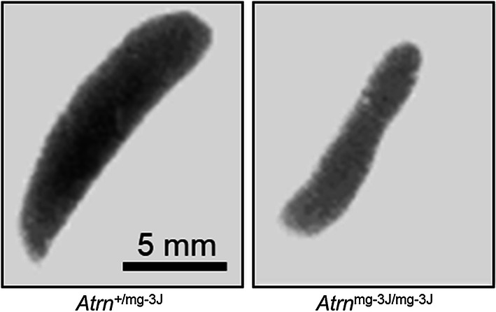
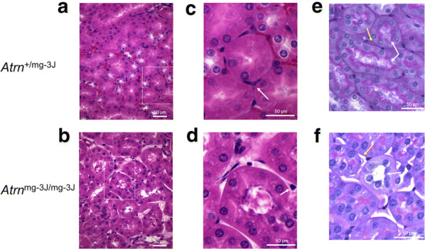
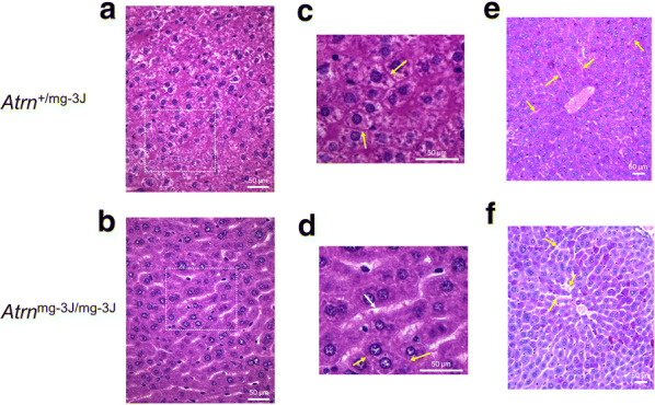
Objective: Neurodegeneration and hair pigmentation alterations in mice occur consequent to aberrations at the Atrn locus coding for the transmembrane form of attractin. Earlier results pointed to a possible involvement in intracellular trafficking/export of secretory vesicles containing proteoglycan. Here we examined kidney and liver, both heavily dependent upon proteoglycan, of attractin-deficient mice to determine whether abnormalities were observed in these tissues.

Results: Histological and histochemical analysis to detect glycosylated protein identified a severe loss in attractin-deficient mice of extracellular proteoglycan between kidney tubules in addition to a loss of glycosylated material within the intratubular brush border. In the liver, extracellular matrix material was significantly depleted between hepatocytes together with swollen sinuses and aberrations in the proteoglycan-dependent space of Disse. These results are consistent with a generalized defect in extracellular proteoglycan deposition in Atrn-mutant mice and support previous reports suggesting a role for attractin in the secretory vesicle pathway.

Keywords: Attractin; Extracellular matrix; Histology; Kidney; Liver; Proteoglycan.


Conflict of interest statement

The authors declare that they have no competing interests.

Figures

Fig. 1
Fig. 1
Spleens of older (~ 5 month) Atrnmg−3J/mg−3J mice (right panel; × 3.5) are consistently half to two-thirds the size of age-matched controls (left panel; ×3.5), reflected also by mononuclear cell counts (wild-type: 6.17 × 107 cells/spleen, Atrn-mutant: 3.9 × 107 cells/spleen; pool of 3 for each genotype)
Fig. 2
Fig. 2
a Normal (Atrn+/mg−3J) kidney (H&E); bAtrnmg−3J/mg−3J kidney (H&E). Note in the normal condition the dense staining basement membrane surrounding each tubule which also forms a tight junction between the tubules. This staining is absent in the Atrn-mutant specimen. c, d Higher resolution images of images within insets (dotted lines) in a, b. Note that in both specimens there are interstitial cells between the tubules (arrows) but only in the normal section are these cells embedded in basement membrane. For all images, results are representative of more than 5 sections from 10 paired sibling comparisons. e Normal kidney; fAtrnmg−3J/mg−3J kidney (PAS stain). The intralumenal brush border of Atrnmg−3J/mg−3J kidney tubules is depleted of extracellular matrix on comparison with control kidney (white arrows), and the intertubular space is similarly depleted (yellow arrows). For all images, results are representative of more than 5 sections from 10 paired sibling comparisons
Fig. 3
Fig. 3
a Normal liver and bAtrnmg−3J/mg−3J liver (H&E stain). The sinusoids (white space between rows of hepatocytes) are readily apparent in the mutant mice. Glycogen does not stain with H&E leading to light areas within the control cells, an effect not seen in the mutant indicating depletion of glycogen. c, d Higher resolution images of images within insets (dotted lines) in a, b. White arrow identifies sinusoid, yellow arrows indicate intercellular space. e Normal liver; fAtrnmg−3J/mg−3J liver (PAS stain). Comparison of sinusoids (yellow arrows) clearly indicates presence of proteoglycan contributing to the perisinusoidal space of Disse in the control liver while such signal is absent in the attractin-deficient liver. Note also (at the yellow arrows) evidence for sinusoidal swelling in the Atrnmg−3J/mg−3J liver accompanied by a more general lack of inter-hepatocyte ECM on comparison with control

References

    1. Duke-Cohan JS, Gu J, McLaughlin DF, Xu Y, Freeman GJ, Schlossman SF. Attractin (DPPT-L), a member of the CUB family of cell adhesion and guidance proteins, is secreted by activated human T lymphocytes and modulates immune cell interactions. Proc Natl Acad Sci USA. 1998;95(19):11336–11341. doi: 10.1073/pnas.95.19.11336. - DOI - PMC - PubMed
    1. Gunn TM, Miller KA, He L, Hyman RW, Davis RW, Azarani A, et al. The mouse mahogany locus encodes a transmembrane form of human attractin. Nature. 1999;398(6723):152–156. doi: 10.1038/18217. - DOI - PubMed
    1. Tang W, Gunn TM, McLaughlin DF, Barsh GS, Schlossman SF, Duke-Cohan JS. Secreted and membrane attractin result from alternative splicing of the human ATRN gene. Proc Natl Acad Sci USA. 2000;97(11):6025–6030. doi: 10.1073/pnas.110139897. - DOI - PMC - PubMed
    1. Azouz A, Gunn TM, Duke-Cohan JS. Juvenile-onset loss of lipid-raft domains in attractin-deficient mice. Exp Cell Res. 2007;313(4):761–771. doi: 10.1016/j.yexcr.2006.11.018. - DOI - PubMed
    1. He L, Gunn TM, Bouley DM, Lu XY, Watson SJ, Schlossman SF, et al. A biochemical function for attractin in agouti-induced pigmentation and obesity. Nat Genet. 2001;27(1):40–47. doi: 10.1038/83741. - DOI - PubMed