. 2020 Jun;8(1):e000189.
doi: 10.1136/jitc-2019-000189. Epub 2020 Jun 23.

CD200 promotes immunosuppression in the pancreatic tumor microenvironment

Affiliations

CD200 promotes immunosuppression in the pancreatic tumor microenvironment

Fouad Choueiry et al. J Immunother Cancer. 2020 Jun.

Abstract

Background: A significant challenge to overcome in pancreatic ductal adenocarcinoma (PDAC) is the profound systemic immunosuppression that renders this disease non-responsive to immunotherapy. Our supporting data provide evidence that CD200, a regulator of myeloid cell activity, is expressed in the PDAC microenvironment. Additionally, myeloid-derived suppressor cells (MDSC) isolated from patients with PDAC express elevated levels of the CD200 receptor (CD200R). Thus, we hypothesize that CD200 expression in the PDAC microenvironment limits responses to immunotherapy by promoting expansion and activity of MDSC.

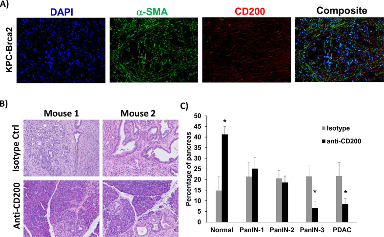
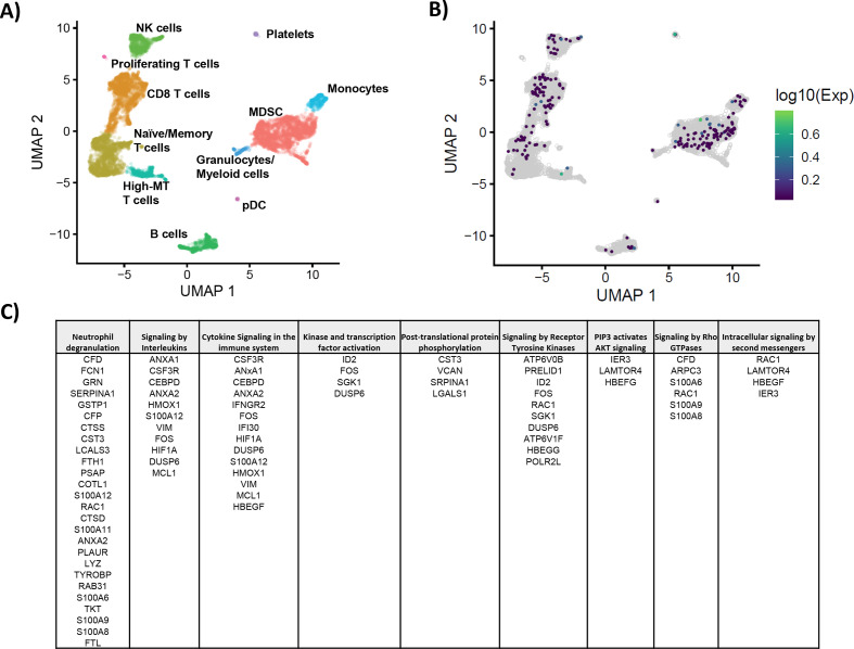
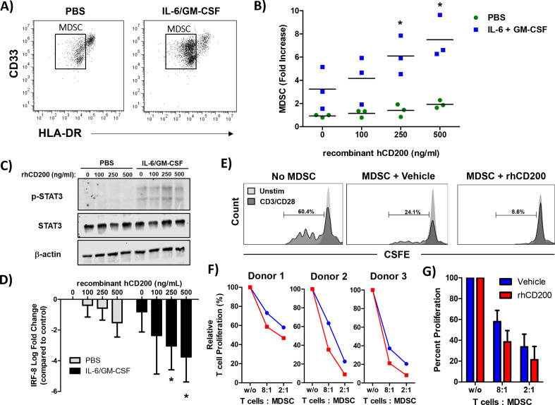
Methods: Immunofluorescent staining was used to determine expression of CD200 in murine and human PDAC tissue. Flow cytometry was utilized to test for CD200R expression by immune populations in patient blood samples. In vivo antibody blocking of CD200 was conducted in subcutaneous MT-5 tumor-bearing mice and in a genetically engineered PDAC model (KPC-Brca2 mice). Peripheral blood mononuclear cells (PBMC) from patients with PDAC were analyzed by single-cell RNA sequencing. MDSC expansion assays were completed using healthy donor PBMC stimulated with IL-6/GM-CSF in the presence of recombinant CD200 protein.

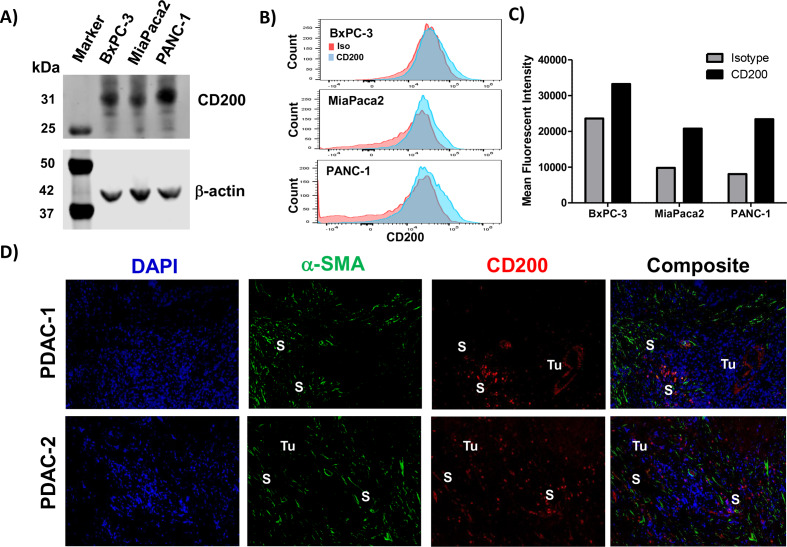
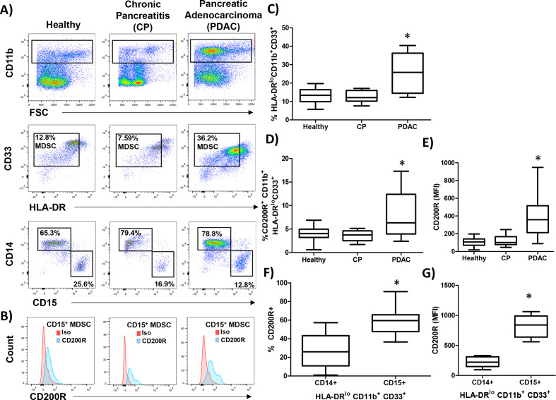
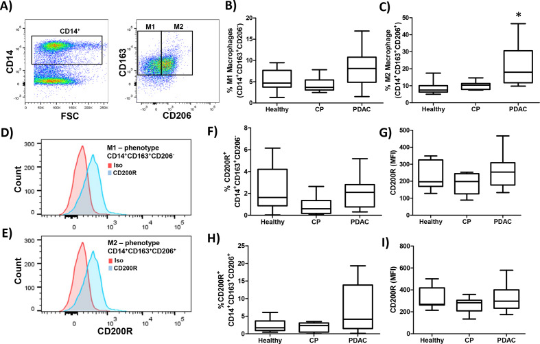
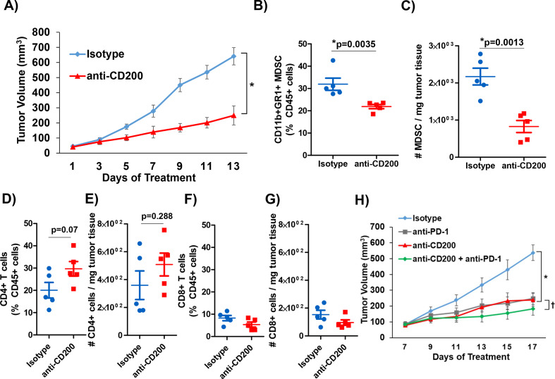
Results: We found expression of CD200 by human pancreatic cell lines (BxPC3, MiaPaca2, and PANC-1) as well as on primary epithelial pancreatic tumor cells and smooth muscle actin+ stromal cells. CD200R expression was found to be elevated on CD11b+CD33+HLA-DRlo/- MDSC immune populations from patients with PDAC (p=0.0106). Higher expression levels of CD200R were observed in CD15+ MDSC compared with CD14+ MDSC (p<0.001). In vivo studies demonstrated that CD200 antibody blockade limited tumor progression in MT-5 subcutaneous tumor-bearing and in KPC-Brca2 mice (p<0.05). The percentage of intratumoral MDSC was significantly reduced in anti-CD200 treated mice compared with controls. Additionally, in vivo blockade of CD200 can also significantly enhance the efficacy of PD-1 checkpoint antibodies compared with single antibody therapies (p<0.05). Single-cell RNA sequencing of PBMC from patients revealed that CD200R+ MDSC expressed genes involved in cytokine signaling and MDSC expansion. Further, in vitro cytokine-driven expansion and the suppressive activity of human MDSC was enhanced when cocultured with recombinant CD200 protein.

Conclusions: These results indicate that CD200 expression in the PDAC microenvironment may regulate MDSC expansion and that targeting CD200 may enhance activity of checkpoint immunotherapy.

Keywords: gastroenterology; immunology; myeloid-derived suppressor cells; tumor microenvironment; tumors.


Conflict of interest statement

Competing interests: None declared.

Figures

Figure 1
Figure 1
Pancreatic tumor and stromal cells express elevated levels of CD200. Cell lysates from pancreatic cancer cell lines (BxPC3, MiaPaca2, PANC-1) were analyzed by (A) immunoblot for CD200 with β-actin as a loading control. (B) CD200 surface staining on the pancreatic cancer cell lines were analyzed by flow cytometry (Red, Isotype control; Blue, CD200). (C) Mean fluorescent intensity values from flow cytometry stained cell lines for CD200. (D) Archived surgical patient PDAC specimens were stained by IF for DAPI (blue), α-SMA (green), and CD200 (red). Tumor (Tu) and stromal (S) positive compartments of the tissue are marked in white. α-SMA-alpha-smooth muscle actin.
Figure 2
Figure 2
CD200 receptor (CD200R) is elevated on MDSC from patients with PDAC. PBMC were isolated from healthy donors (n=9), patients with chronic pancreatitis (CP; n=10), or pancreatic ductal adenocarcinoma (PDAC; n=17) and (A) stained by flow cytometry for granulocytic (CD11b+CD33+HL-DR−/lowCD15+) and monocytic (CD11b+CD33+HL-DR-/lowCD14+) MDSC. (B) Representative CD200R (blue) or isotype control (red) staining of CD15+ MDSC. (C) Percent total MDSC, (D) CD200R positive cells, and (E) mean fluorescent intensity (MFI) were quantified across all patient groups. (F) Percent and (G) MFI of either CD14+ or CD15+ MDSC that express CD200R. Mean±SD; *p<0.05. MDSC, myeloid-derived suppressor cells; PBMC, peripheral blood mononuclear cells.
Figure 3
Figure 3
No significant increase in expression of CD200R on M2 macrophages in patients with PDAC. PBMC were isolated from healthy donors (n=9), patients with CP (n=10), or PDAC (n=11) and (A) stained by flow cytometry for M1 (CD14+CD163+CD206) or M2 (CD14+CD163+CD206+) macrophages. Percent of (B) M1 or (C) M2 macrophages were quantified across the patient groups. Representative CD200R (blue) or isotype control (red) staining of (D) M1 or (E) M2 macrophages. (F) Percent and (G) MFI of M1-like macrophages expressing CD200R quantified across all patient groups. (H) Percent and (I) MFI of M2-like macrophages expressing CD200R quantified across all patient groups. Mean±SD; *p<0.05. CP, chronic pancreatitis; MDSC, myeloid-derived suppressor cells; MFI, mean fluorescent intensity; PBMC, peripheral blood mononuclear cells; PDAC, pancreatic ductal adenocarcinoma.
Figure 4
Figure 4
CD200 antibody blockade elicits antitumor response. KPC-derived MT5 tumor were subcutaneously injected into C57BL/6 mice. (A) Mice were treated with 200 µg/mouse of CD200 or isotype control antibodies 3× a week (n=5 mice/group). Intratumoral flow cytometry staining (percent of CD45+ cells and number per mg of tumor tissue) for (B) and (C) MDSC (CD11b+GR1+), (D) and (E) CD4+ T cells, (F) and (G) CD8+ T cells. (H) C57BL/6 mice were inoculated subcutaneously with MT5 tumor cells and were treated once palpable. MT5 tumor-bearing mice were treated with 200 µg/mouse of anti-CD200, PD-1, or isotype control antibodies 3× a week (n=5 mice/group). Mean±SD; * and †=p<0.05. MDSC, myeloid-derived suppressor cells.
Figure 5
Figure 5
CD200 antibody blockade limits tumor progression in GEM model of PDAC. (A) Representative immunofluorescence from pancreatic tissue of KPC-Brca2 mice stained for DAPI (blue), a-SMA (green), and CD200 (red). (B) KPC-Brca2 mice were treated at 6 weeks of age with 200 µg (intraperitoneal injection three times/week) of isotype control or CD200 antibodies (n=5 mice/group). (C) Histology was pathologically scored for PanIN lesions and quantified. Mean±SD; *p<0.05. GEM, genetically engineered mouse; PDAC, pancreatic ductal adenocarcinoma; α-SMA-alpha-smooth muscle actin.
Figure 6
Figure 6
Analysis of genes expressed by CD200R+ populations from PBMC of patients with PDAC. PBMC from patients with PDAC (n=3) were processed by single-cell RNA sequencing and analyzed by Chromium 10× genomics. (A) All three patients were aggregated and clustered into different immune populations based on gene expression data. (B) Cells overexpressing genes involved in CD200R signaling (CD200R, Dok1, and Dok2) were identified by taking the maximum of the scaled, size-factor normalized expression values for these genes in each cell. (C) Reactome Pathway Profile software was used to analyze the significant interactions of genes that were expressed by CD200R+ MDSC. The genes that were overexpressed by CD200R+ MDSC were analyzed by the Reactome software to determine potential pathways that may be active in CD200R expressing cells. MDSC, myeloid-derived suppressor cells; PBMC, peripheral blood mononuclear cells; PDAC, pancreatic ductal adenocarcinoma; UMAP, Uniform Manifold Approximation and Projection.
Figure 7
Figure 7
CD200 enhances the cytokine-driven differentiation and suppressive activity of MDSC in vitro. Normal donor PBMC were cultured for 7 days with 10 ng/mL of IL-6 and 10 ng/mL of GM-CSF and stained by flow cytometry for the percentage of MDSC (CD11b+CD33+HLA-DRlo). (A) Representative flow cytometry dot plots from unstimulated or IL-6/GM-CSF stimulated PBMC after 7 days of differentiation. (B) During differentiation, cells were cultured in the presence of recombinant human CD200 protein (rhCD200). (C) Healthy donor PBMC were stimulated with 10 ng/mL of IL-6 and GM-CSF for 30 min with increasing concentrations of rhCD200 protein. Cell lysates were analyzed for STAT3 phosphorylation (p-STAT3) with β-actin as a loading control. (D) Healthy donor PBMC were stimulated with 10 ng/mL of IL-6 and GM-CSF with increasing concentrations of rhCD200 protein for 2 hours. RNA was isolated and expression of IRF-8 was analyzed by real-time PCR. (E) PBMC from healthy donor blood was stimulated with 10 ng/mL of IL-6 and GM-CSF to differentiate cells into MDSC for 5 days. Cells were then cultured with vehicle control or 200 ng/mL rhCD200 for 48 hours. MDSC were then cocultured with autologous CFSE-labeled T cells stimulated. T cells were stimulated with CD3/CD28 beads and proliferation was measured after 4 days by CFSE dilution. (F) Individual donor T-cell proliferation and (G) quantification across all donors. Mean±SD; *p<0.05. MDSC, myeloid-derived suppressor cells; PBMC, peripheral blood mononuclear cells.

References

    1. Von Hoff DD, Ervin T, Arena FP, et al. . Increased survival in pancreatic cancer with nab-paclitaxel plus gemcitabine. N Engl J Med 2013;369:1691–703. 10.1056/NEJMoa1304369 - DOI - PMC - PubMed
    1. Koutsounas I, Giaginis C, Patsouris E, et al. . Current evidence for histone deacetylase inhibitors in pancreatic cancer. World J Gastroenterol 2013;19:813–28. 10.3748/wjg.v19.i6.813 - DOI - PMC - PubMed
    1. Azar I, Virk G, Esfandiarifard S, et al. . Treatment and survival rates of stage IV pancreatic cancer at Va hospitals: a nation-wide study. J Gastrointest Oncol 2019;10:703–11. 10.21037/jgo.2018.07.08 - DOI - PMC - PubMed
    1. Siegel RL, Miller KD, Jemal A. Cancer statistics, 2019. CA Cancer J Clin 2019;69:7–34. 10.3322/caac.21551 - DOI - PubMed
    1. Kneuertz PJ, Cunningham SC, Cameron JL, et al. . Palliative surgical management of patients with unresectable pancreatic adenocarcinoma: trends and lessons learned from a large, single institution experience. J Gastrointest Surg 2011;15:1917–27. 10.1007/s11605-011-1665-9 - DOI - PMC - PubMed

Publication types

MeSH terms