. 2020 Jun;8(1):e000182.
doi: 10.1136/jitc-2019-000182.

Involvement of the M-CSF/IL-34/CSF-1R pathway in malignant pleural mesothelioma

Affiliations

Involvement of the M-CSF/IL-34/CSF-1R pathway in malignant pleural mesothelioma

Thibaut Blondy et al. J Immunother Cancer. 2020 Jun.

Abstract

Background: Malignant pleural mesothelioma (MPM) is a rare and aggressive cancer related to asbestos exposure. The tumor microenvironment content, particularly the presence of macrophages, was described as crucial for the development of the disease. This work aimed at studying the involvement of the M-CSF (CSF-1)/IL-34/CSF-1R pathway in the formation of macrophages in MPM, using samples from patients.

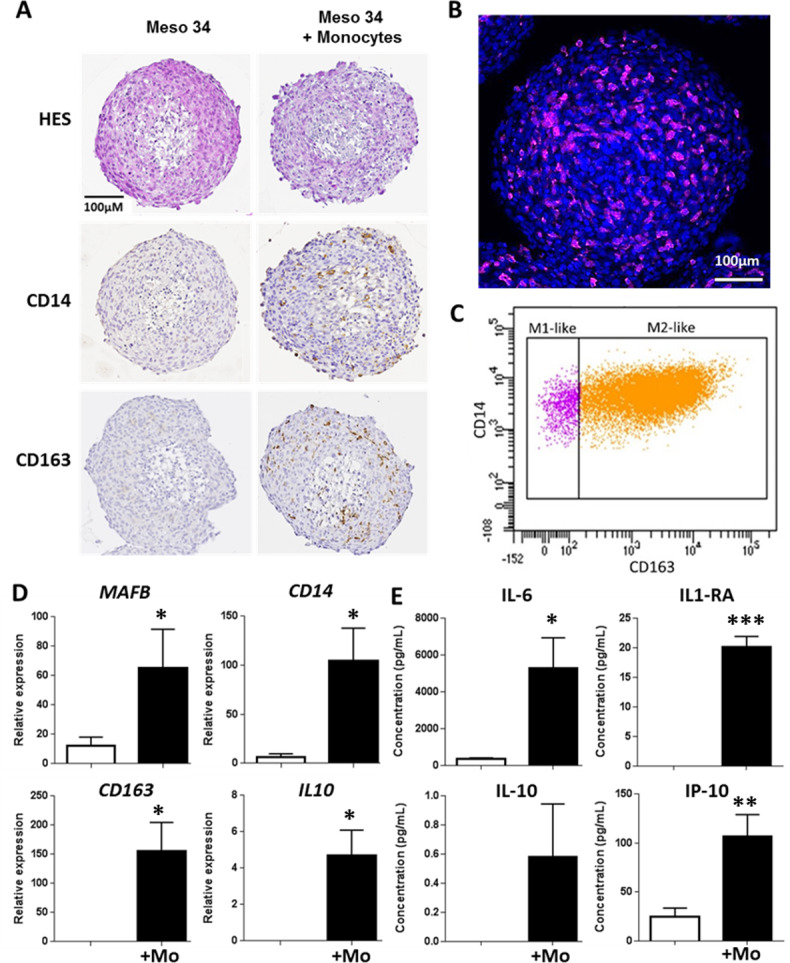
Methods: Pleural effusions (PEs), frozen tumors, primary MPM cells and MPM cell lines used in this study belong to biocollections associated with clinical databases. Cytokine expressions were studied using real-time PCR and ELISA. The Cancer Genome Atlas database was used to confirm our results on an independent cohort. An original three-dimensional (3D) coculture model including MPM cells, monocytes from healthy donors and a tumor antigen-specific cytotoxic CD8 T cell clone was used.

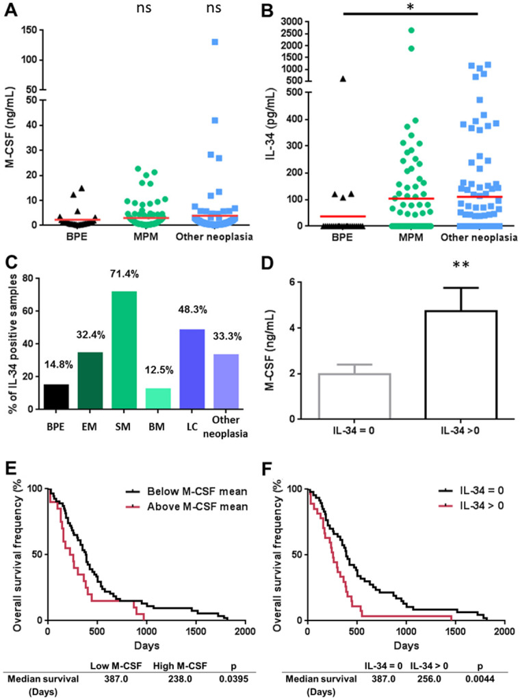
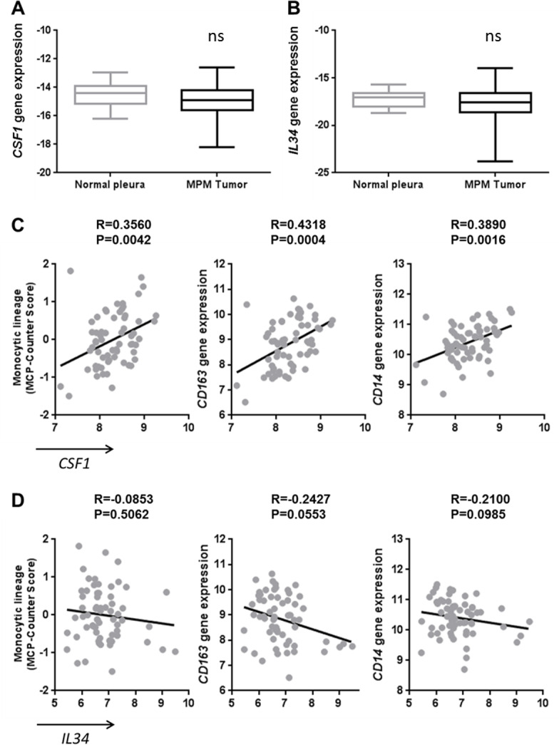
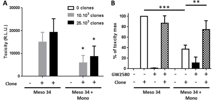
Results: We observed that high interleukin (IL)-34 levels in PE were significantly associated with a shorter survival of patients. In tumors, expression of CSF1 was correlated with 'M2-like macrophages' markers, whereas this was not the case with IL34 expression, suggesting two distinct modes of action of these cytokines. Expression of IL34 was higher in MPM cells compared with primary mesothelial cells. Particularly, high expression of IL34 was observed in MPM cells with an alteration of CDKN2A. Finally, using 3D coculture model, we demonstrated the direct involvement of MPM cells in the formation of immunosuppressive macrophages, through activation of the colony stimulating factor-1 receptor (CSF1-R) pathway, causing the inhibition of cytotoxicity of tumor antigen-specific CD8+ T cells.

Conclusions: The M-CSF/IL-34/CSF-1R pathway seems strongly implicated in MPM and could constitute a therapeutic target to act on immunosuppression and to support immunotherapeutic strategies.

Keywords: immunology; oncology; tumors.


Conflict of interest statement

Competing interests: None declared.

Figures

Figure 1
Figure 1
Expression of M-CSF and IL-34 in pleural effusions from patients and prognostic value. (A) M-CSF and (B) IL-34 expression in pleural effusions from patients with MPM (n=96), other neoplasia (n=105) or BPE (n=26). Red bars correspond to means. (C) Percentage of IL-34 positive samples in the different groups of patients. (D) Levels of M-CSF in IL-34 negative and positive samples. Patients were split in ‘high expression’ and ‘low expression’ groups based on the mean of expression of M-CSF (E) or on positive and negative expression of IL-34 (F) in MPM pleural effusions, and differences in survival between the two groups were assessed using log-rank tests. *p<0.05; **p<0.01. BM, biphasic mesothelioma; BPE, benign pleural effusion; EM, epithelioid mesothelioma; LC, lung cancer; MPM, malignant pleural mesothelioma; SM, sarcomatoid mesothelioma.
Figure 2
Figure 2
Correlation of CSF1 expression with tumor-associated macrophage markers in malignant pleural mesothelioma (MPM) tumors. (A) CSF1 and (B) IL34 gene expressions measured using quantitative real-time PCR in MPM tumors (n=178) and normal pleura (n=26). (C–D) Correlation between CSF1 (C) or IL34 (D) expressions and monocytic lineage-specific CD163 and CD14 expressions using transcriptomic data of MPM tumor samples (n=63).
Figure 3
Figure 3
Expression of CSF1 and IL34 in MPM cells. (A) CSF1 and (B) IL34 gene expressions measured using quantitative real-time PCR (qRT-PCR) in primary malignant pleural mesothelioma (MPM) cells (n=69) and normal mesothelial cells (n=4; MC). (C) CSF1 and (D) IL34 gene expressions measured using qRT-PCR in primary MPM cells with or without CDKN2A genetic alteration. (E) CSF1 and (F) IL34 gene expressions measured using qRT-PCR in primary MPM cells with or without NF2 genetic alteration. *p<0.05; ***p<0.001.
Figure 4
Figure 4
Evaluation of malignant pleural mesothelioma (MPM) cell capacity to drive monocyte differentiation into M2-like macrophages using a multicellular tumor spheroid (MCTS) model. Meso 34 cells were cultured with or without 30% of monocytes (Mo) in low adherence conditions for 3 days. (A) Macrophage phenotype was studied using immunohistochemistry with CD14 or CD163 labeling. To confirm the presence of macrophages, (B) MCTSs were labeled with an anti-CD163 antibody coupled to Alexa-Fluor 647 (purple), Hoechst for nuclei staining (blue) and observed using confocal microscopy. (C) Representative analysis of three independent flow cytometry experiments showing CD14 and CD163 expression on HLA-DR+ and CD14+ cells in MCTS. (D) Expressions of MAFB, CD14, CD163 and IL10 mRNA were measured using quantitative real-time PCR. Results are means±SEM of six independent experiments. *p<0.05; **p<0.01. (E) Impact of the presence of macrophages in MCTS was determined by measuring the levels of IL-6, IL-1RA, IL-10 and IP-10 (CXCL10) in MCTS culture supernatants. Results are means±SEM of six independent experiments. **p<0.01.
Figure 5
Figure 5
Effect of CSF-1R inhibition on macrophages in multicellular tumor spheroid (MCTS). Meso 34 cells were cultured with or without 30% of monocytes (Mono) in low adherence conditions for 3 days in the presence or not of 1 µM GW2580. (A) Expressions of MAFB, CD14, CD163 and IL10 mRNA were measured using quantitative real-time PCR. Results are means±SEM of three independent experiments. *p<0.05; ns, non-significant. (B) Levels of interleukin (IL)-6, IL-1RA, IL-1β, tumor necrosis factor alpha (TNFα), IL-10 and IP-10 (CXCL10) in MCTS culture supernatants. Results are means±SEM of six independent experiments. *p<0.05; **p<0.01; ns, non-significant.
Figure 6
Figure 6
Effect of macrophage and CSF-1R inhibition on the specific CD8 T cell clone cytotoxic activity against MPM cells. Meso 34 NanoLuc cells were cultured with or without monocytes (Mono) in low adherence conditions. After 3 days, a MUC1-specific CD8 T cell clone was added for 24 hours. Then, supernatants were collected and NanoLuc activity was measured to determine cell lysis. (A) Effect of the presence of macrophages on the cytotoxic activity of the MUC1-specific T cell clone. Results are means±SEM of four independent experiments. Meso34 vs Meso34+Mono: *p<0.05. (B) Impact of the inhibition of CSF-1R, using GW2580 (1 µM), on the T cell clone cytotoxic activity. **p<0.01; ***p<0.001. CSF-1R, colony stimulatingfactor-1 Receptor; MPM, malignant pleural mesothelioma; RLU, relative light units.

References

    1. Vogelzang NJ, Rusthoven JJ, Symanowski J, et al. . Phase III study of pemetrexed in combination with cisplatin versus cisplatin alone in patients with malignant pleural mesothelioma. J Clin Oncol 2003;21:2636–44. 10.1200/JCO.2003.11.136 - DOI - PubMed
    1. Burt BM, Rodig SJ, Tilleman TR, et al. . Circulating and tumor-infiltrating myeloid cells predict survival in human pleural mesothelioma. Cancer 2011;117:5234–44. 10.1002/cncr.26143 - DOI - PubMed
    1. Ujiie H, Kadota K, Nitadori J-I, et al. . The tumoral and stromal immune microenvironment in malignant pleural mesothelioma: a comprehensive analysis reveals prognostic immune markers. Oncoimmunology 2015;4:e1009285. 10.1080/2162402X.2015.1009285 - DOI - PMC - PubMed
    1. Mantovani A, Marchesi F, Malesci A, et al. . Tumour-Associated macrophages as treatment targets in oncology. Nat Rev Clin Oncol 2017;14:399–416. 10.1038/nrclinonc.2016.217 - DOI - PMC - PubMed
    1. Cornelissen R, Lievense LA, Maat AP, et al. . Ratio of intratumoral macrophage phenotypes is a prognostic factor in epithelioid malignant pleural mesothelioma. PLoS One 2014;9:e106742. 10.1371/journal.pone.0106742 - DOI - PMC - PubMed

Publication types

MeSH terms