. 2020 Aug 17;94(17):e00231-20.
doi: 10.1128/JVI.00231-20. Print 2020 Aug 17.

Induction of Rod-Shaped Structures by Herpes Simplex Virus Glycoprotein I

Affiliations

Induction of Rod-Shaped Structures by Herpes Simplex Virus Glycoprotein I

Wuchao Zhang et al. J Virol. .

Abstract

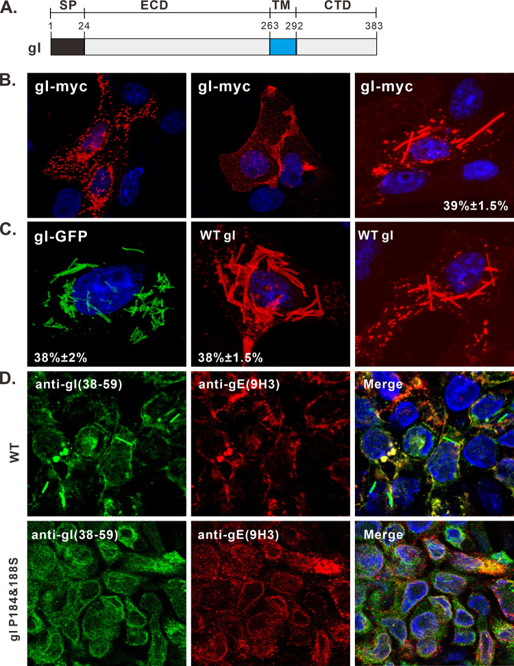
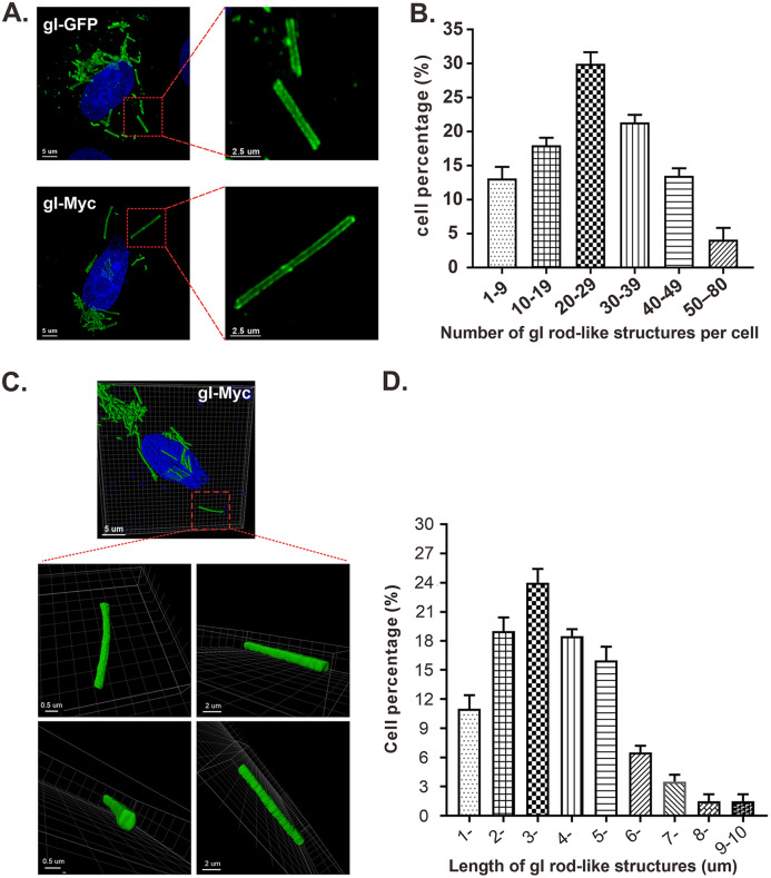
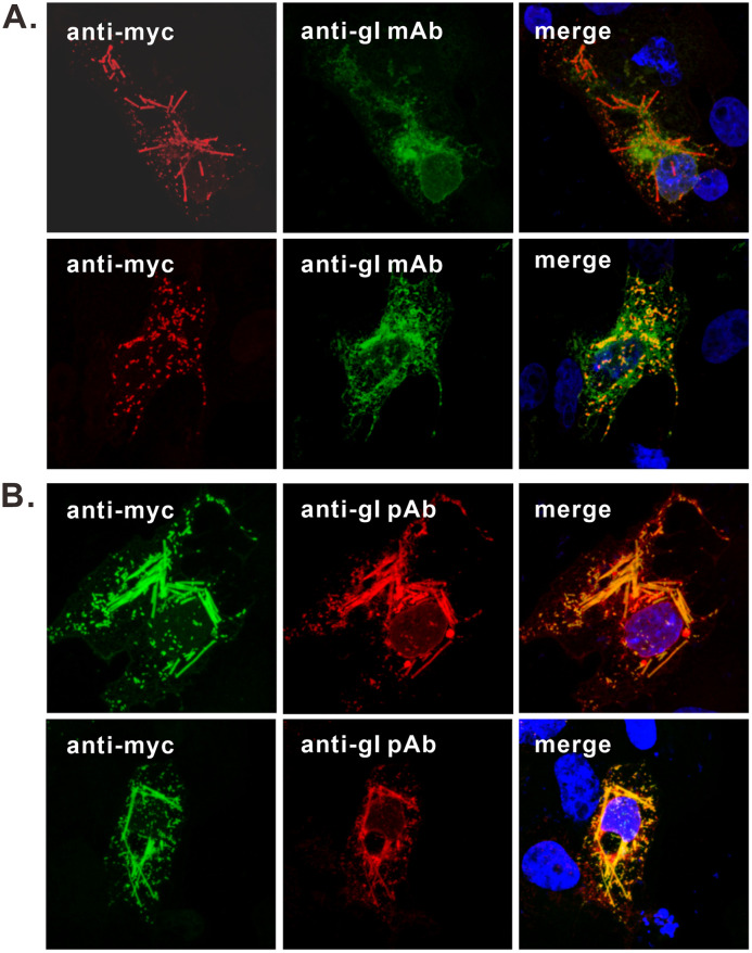
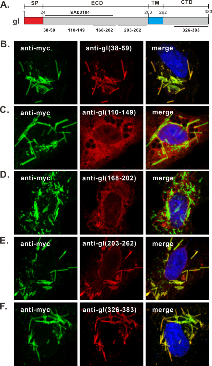
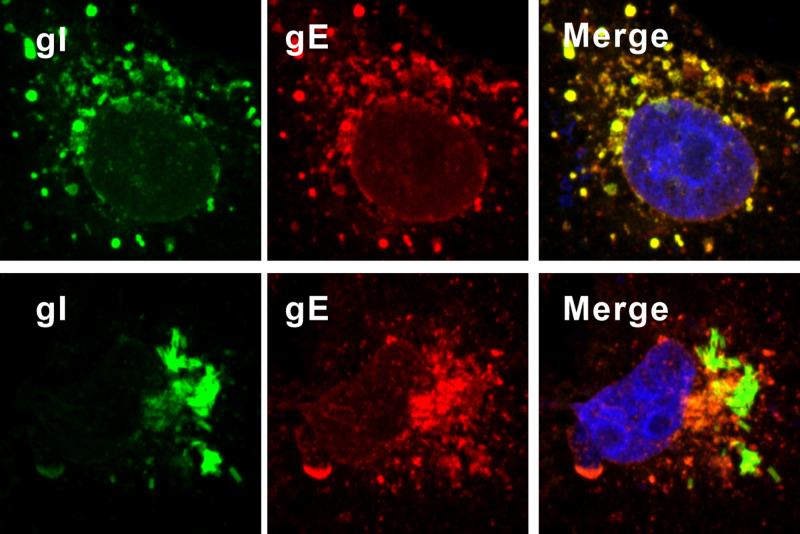
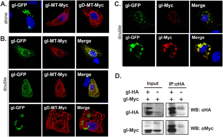
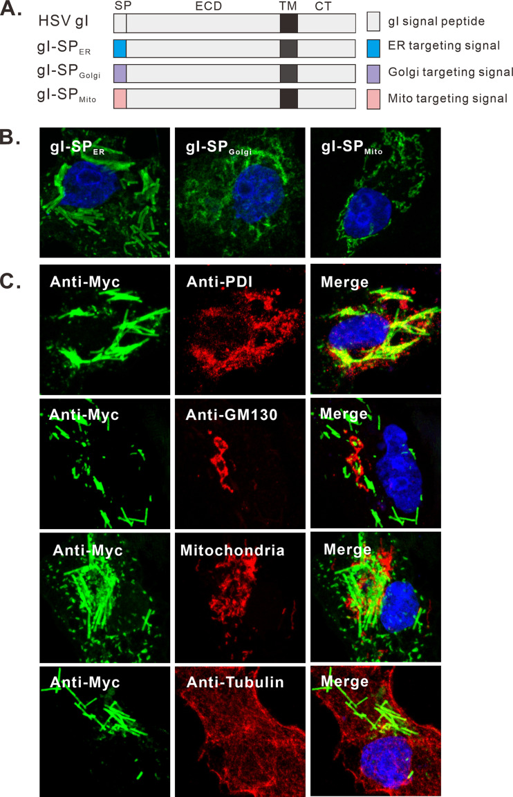
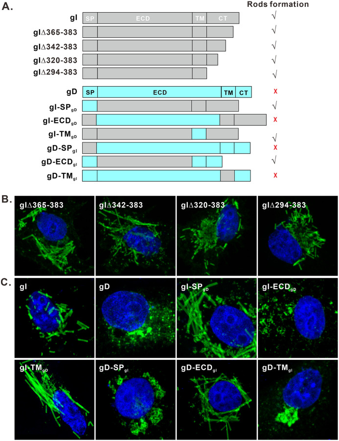
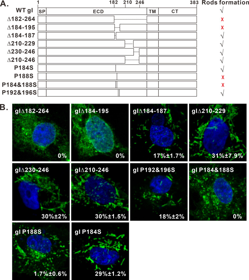
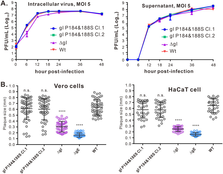
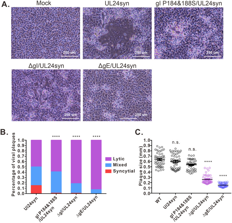
The envelope glycoprotein I (gI) of herpes simplex virus 1 (HSV-1) is a critical mediator of virus-induced cell-to-cell spread and cell-cell fusion. Here, we report a previously unrecognized property of this molecule. In transfected cells, the HSV-1 gI was discovered to induce rod-shaped structures that were uniform in width but variable in length. Moreover, the gI within these structures was conformationally different from the typical form of gI, as a previously used monoclonal antibody mAb3104 and a newly made peptide antibody to the gI extracellular domain (ECD) (amino acids [aa] 110 to 202) both failed to stain the long rod-shaped structures, suggesting the formation of a higher-order form. Consistent with this observation, we found that gI could self-interact and that the rod-shaped structures failed to recognize glycoprotein E, the well-known binding partner of gI. Further analyses by deletion mutagenesis and construction of chimeric mutants between gI and gD revealed that the gI ECD is the critical determinant, whereas the transmembrane domain served merely as an anchor. The critical amino acids were subsequently mapped to proline residues 184 and 188 within a conserved PXXXP motif. Reverse genetics analyses showed that the ability to induce a rod-shaped structure was not required for viral replication and spread in cell culture but rather correlated positively with the capability of the virus to induce cell fusion in the UL24syn background. Together, this work discovered a novel feature of HSV-1 gI that may have important implications in understanding gI function in viral spread and pathogenesis.IMPORTANCE The HSV-1 gI is required for viral cell-to-cell spread within the host, but the molecular mechanisms of how gI exactly works have remained poorly understood. Here, we report a novel property of this molecule, namely, induction of rod-shaped structures, which appeared to represent a higher-order form of gI. We further mapped the critical residues and showed that the ability of gI to induce rod-shaped structures correlated well with the capability of HSV-1 to induce cell fusion in the UL24syn background, suggesting that the two events may have an intrinsic link. Our results shed light on the biological properties of HSV-1 gI and may have important implications in understanding viral pathogenesis.

Keywords: cell-cell fusion; cell-to-cell spread; glycoprotein I (gI); herpes simplex virus; membrane tubulation; rod-shaped structures.


Figures

FIG 1
FIG 1
Induction of rod-shaped structures by HSV-1 gI. (A) Diagram of domain organization of HSV-1 KOS strain glycoprotein gI. (B and C) Vero cells were transfected to express HSV-1 gI-Myc, gI-GFP, and untagged gI. At 18 to 24 h posttransfection, the cells were fixed, permeabilized, and revealed by antibodies specific for the Myc tag, gI antigen, and HA tag or by GFP fluorescence. DAPI was used to stain cell nuclei (blue). The representative images were captured with a Leica confocal microscope and processed by using ImageJ. The percentage of cells showing rod-shaped structures relative to total gI-positive cells is also shown in the pictures. (D) HaCaT cells grown on coverslips were infected with WT HSV or the mutant gI P184&188S at an MOI of 0.1 at 37°C. After 1 h of incubation, the cells were rinsed with PBS and supplemented with fresh infection medium. At 24 h postinfection, the cells were fixed and stained with rabbit polyclonal antibodies against gI and mouse monoclonal antibodies against gE (red) as indicated. The rest of the steps were the same as panels B and C.
FIG 2
FIG 2
Analysis of the number, size, and dimensions of HSV-1 gI rod-shaped structures. Vero cells were transfected to express gI-Myc or gI-GFP. At 18 to 24 h posttransfection, the cells were fixed, permeabilized, and stained with antibodies to Myc epitope tag (for cells expressing gI-myc). DAPI was used to stain cell nuclei (blue). The representative images were captured with a Leica confocal microscope and processed by using ImageJ and Imaris. (A) Images of gI-GFP and gI-myc at higher magnification. (B) Statistical analysis of the number per cell of gI rods. The y axis indicates the percentage of cells having rod structures, and the x axis indicates the number range of rods per cell. (C) Three-dimensional reconstruction of gI rod structures by Imaris software. The size bars are indicated. (D) Quantification analysis of the length of rod structures. The y axis indicates the percentage of cells having rod structures, and the x axis indicates the length range of rods.
FIG 3
FIG 3
The gI within the rod-shaped structures is structurally different from the typical form of gI. Vero cells were transfected to express gI-Myc. At 18 to 24 h posttransfection, the cells were fixed, permeabilized, and doubly stained with antibodies to Myc epitope tag and gI monoclonal antibody mAb3104 (A) or rabbit polyclonal antibodies to gI extracellular domain (UP1725) (B). DAPI was used to stain cell nuclei (blue). The representative images were captured with a Leica confocal microscope and processed using ImageJ.
FIG 4
FIG 4
The gI region aa 110 to 202 in the rod-shaped structures is not exposed outside. (A) Diagram of gI showing the recognition sites of gI antibodies. (B to F) Vero cells were transfected to express gI-Myc. At 18 to 24 h posttransfection, the cells were fixed, permeabilized, and doubly stained with antibodies specific for Myc tag (green) and gI peptide antibodies (red) as indicated. DAPI was used to stain cell nuclei (blue). The representative images were captured with a Leica confocal microscope and processed using ImageJ.
FIG 5
FIG 5
The gI within rod-shaped structures does not colocalize gE. Vero cells were transfected to coexpress gI-myc and gE-HA. At 18 to 24 h posttransfection, the cells were fixed, permeabilized, and costained with antibodies specific for Myc tag (green) and HA antigen (red). The representative images were captured with a Leica confocal microscope and processed using ImageJ.
FIG 6
FIG 6
Analysis of HSV-1 gI self-interaction. Vero cells were transfected to either singly express (A) or coexpress gI-GFP and gI-MT-myc, or gD-MT-myc (B) or gI-myc (C). The singly expressed proteins served as a control. At 18 to 24 h posttransfection, the cells were fixed, permeabilized, and stained with antibodies specific for Myc tag (red). The representative images were captured with a Leica confocal microscope and processed by using ImageJ. (D) Vero cells were transfected to express gI-HA and gI-myc or to singly express gI-myc. At 24 h posttransfection, the cells were lysed, and gI was immunoprecipitated with antibodies to HA epitope. The proteins bounded to the beads were separated by SDS-PAGE, followed by Western blotting with antibodies to either HA or c-myc epitope.
FIG 7
FIG 7
Analysis of the membrane origin of gI rod-shaped structures. (A) Schematic diagrams of chimeric gI-myc constructs in which the gI signal peptide was replaced with either ER, Golgi, or mitochondrial targeting signal. (B) Vero cells were transfected to express the gI mutants and stained with antibodies to Myc epitope. (C) Vero cells were transfected to express gI-Myc. At 18 to 24 h posttransfection, the cells were fixed, permeabilized, and costained with antibodies to Myc tag (green) and other subcellular markers (red). The cell nuclei were stained with DAPI (blue). The representative images were captured with a Leica confocal microscope and processed by using ImageJ.
FIG 8
FIG 8
The gI extracellular domain is the critical determinant for inducing rod-shaped structures. (A) Diagram of gI/gD chimeric mutants and gI CT truncated mutants. The corresponding SP, ECD, and TM regions were swapped in a reciprocal manner. (B and C) Mutants were transiently expressed in Vero cells and were with antibodies to Myc and HA epitopes, followed with Alexa Fluor 488-conjugated secondary antibodies. The cell nuclei were stained with DAPI (blue). The representative images were captured with a Leica confocal microscope and processed by using ImageJ.
FIG 9
FIG 9
Mapping the key residues for formation of the gI rod-shaped structures. (A) Mutagenesis analysis of the gI extracellular domain. The deleted region is shown as a dashed line and point mutation as a vertical line with the relative positions indicated in the name of individual mutants. The capacity of inducing rod-shaped structures for each mutant is shown on the right. √, capable; ×, incapable. (B) Immunofluorescence staining of some of the mutants in transfected Vero cells with antibodies to Myc epitope. The images were obtained by confocal microscopy.
FIG 10
FIG 10
Effect of gI P184&188S mutation on viral replication and spread in cell culture. (A) Growth curve of WT HSV-1 and gI mutants in HaCaT cells at an MOI of 5. The intracellular virus and supernatant were harvested at indicated time points for titer determination by standard viral plaque assay on Vero cells. The data shown are representative of results from three independent experiments. (B) Quantitative analysis of the plaque sizes in Vero and HaCaT cells. Vero (left) and HaCaT (right) cells were infected with HSV-1 WT and recombinant viruses under plaque assay conditions. The cells were fixed at 5 days postinfection and plaques were visualized by staining with crystal violet. The diameters of 50 (Vero cells, left) or 30 (HaCaT cells, right) single plaques for each of the indicated viruses were measured. The horizontal bars indicate the mean for each group. ****, P < 0.001; n.s., not significant.
FIG 11
FIG 11
Effect of gI P184&188S mutations on syncytium formation in the UL24syn background. (A) Virus-induced CPE. Vero cells were infected by the virus as indicated at an MOI of 0.01. At 36 hpi, the virus-induced CPE was taken by a Nikon inverted microscope. The data showed the representative pictures. (B) Quantitative analysis of virus induced-syncytium. Vero cells were infected at an MOI of 0.01. After 1 h of incubation, the cells were rinsed with PBS overlaid with DMEM containing 0.6% methylcellulose and incubated for 36 h. Afterward, the cells were fixed and stained with antibody to VP5, and the cell nuclei were stained with DAPI. The infection focus was scored as syncytial, mixed, or lytic according to the methods in Materials and Methods. (C) Quantitative analysis of the plaque sizes in Vero cells. The diameters of 50 single plaques for each of the indicated viruses were measured. The horizontal bars indicate the mean for each group. ****, P < 0.001; n.s., not significant.

Similar articles

Cited by

References

    1. Davison AJ, Eberle R, Ehlers B, Hayward GS, McGeoch DJ, Minson AC, Pellett PE, Roizman B, Studdert MJ, Thiry E. 2009. The order Herpesvirales. Arch Virol 154:171–177. doi:10.1007/s00705-008-0278-4. - DOI - PMC - PubMed
    1. James SH, Kimberlin DW. 2015. Neonatal herpes simplex virus infection. Infect Dis Clin North Am 29:391–400. doi:10.1016/j.idc.2015.05.001. - DOI - PubMed
    1. Stahl JP, Mailles A. 2019. Herpes simplex virus encephalitis update. Curr Opin Infect Dis 32:239–243. doi:10.1097/QCO.0000000000000554. - DOI - PubMed
    1. Groves MJ. 2016. Genital herpes: a review. Am Fam Physician 93:928–934. - PubMed
    1. Harris SA, Harris EA. 2018. Molecular mechanisms for herpes simplex virus type 1 pathogenesis in Alzheimer's disease. Front Aging Neurosci 10:48. doi:10.3389/fnagi.2018.00048. - DOI - PMC - PubMed

Publication types

LinkOut - more resources