. 2020 Aug 17;94(17):e00833-20.
doi: 10.1128/JVI.00833-20. Print 2020 Aug 17.

Residues within the Foot-and-Mouth Disease Virus 3Dpol Nuclear Localization Signal Affect Polymerase Fidelity

Affiliations

Residues within the Foot-and-Mouth Disease Virus 3Dpol Nuclear Localization Signal Affect Polymerase Fidelity

Anna Kloc et al. J Virol. .

Abstract

Many RNA viruses encode a proof-reading deficient, low-fidelity RNA-dependent polymerase (RdRp), which generates genetically diverse populations that can adapt to changing environments and thwart antiviral therapies. 3Dpol, the RdRp of the foot-and-mouth disease virus (FMDV), is responsible for replication of viral genomes. The 3Dpol N terminus encodes a nuclear localization signal (NLS) sequence,MRKTKLAPT, important for import of the protein to host nucleus. Previous studies showed that substitutions at residues 18 and 20 of the NLS are defective in proper incorporation of nucleotides and RNA binding. Here, we use a systematic alanine scanning mutagenesis approach to understand the role of individual residues of the NLS in nuclear localization and nucleotide incorporation activities of 3Dpol We identify two residues of 3Dpol NLS, T19 and L21, that are important for the maintenance of enzyme fidelity. The 3Dpol NLS alanine substitutions of T19 and L21 results in aberrant incorporation of nucleoside analogs, conferring a low fidelity phenotype of the enzyme. A molecular dynamics simulation of RNA- and mutagen (RTP)-bound 3Dpol revealed that the T19 residue participates in a hydrogen bond network, including D165 in motif F and R416 at the C terminus of the FMDV 3Dpol and RNA template-primer. Based on these findings and previous studies, we conclude that at least the first six residues of theMRKTKLAPT sequence motif play a vital role in the maintenance of faithful RNA synthesis activity (fidelity) of FMDV 3Dpol, suggesting that the role of the NLS motif in similar viral polymerases needs to be revisited.IMPORTANCE In this study, we employed genetic and molecular dynamics approaches to analyze the role of individual amino acids of the FMDV 3Dpol nuclear localization signal (NLS). The NLS residues were mutated to alanine using a type A full-genome cDNA clone, and the virus progeny was analyzed for defects in growth and in competition with the parental virus. We identified two mutants in 3Dpol, T19A and L21A, that exhibited high rate of mutation, were sensitive to nucleotide analogs, and displayed reduced replicative fitness compared to the parental virus. Using molecular dynamics simulation, we demonstrated that residues T19 and L21 played a role in the structural configuration of the interaction network at the 3Dpol palm subdomain. Cumulatively, our data suggest that the T19 and L21 3Dpol amino acids are important for maintaining the fidelity of the FMDV polymerase and ensuring faithful replication of the FMDV genome.

Keywords: 3Dpol; FMDV; RNA-dependent RNA polymerase fidelity; enzyme nuclear localization signal; foot-and-mouth disease virus; picornavirus.


Figures

FIG 1
FIG 1
Schematic representation of the NLS alanine mutagenesis scheme. (A) Image of the FMDV genome with 3Dpol NLS residues, spanning amino acids 16 through 24. (B) Individual amino acids of the 3Dpol NLS mutated to alanine are shown on the left-hand site of the panel, and the alanine mutated nucleotides of each amino acids are marked in red. For reference, the original 3Dpol NLS sequence is indicated at the top of the panel. Plaque assay images of the alanine substitution viruses are shown on the right side of the panel. *, The T24A-induced mutation (GCT) reverted to threonine (ACT).
FIG 2
FIG 2
Analysis of growth kinetics of the 3D alanine mutants. BHK-21 cells were infected with each of the viruses at an MOI of 1 in triplicates. The time points were 0, 6, 8, 24, and 48 hpi, and a plaque assay was used to determine the viral titer at each time point. The error bars represent statistical deviations within the three replicates.
FIG 3
FIG 3
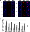
Immunofluorescence analysis of the localization of the 3D protein of the 3D NLS alanine substitution viruses to host nucleus. (A) BHK-21 cells were infected with WT, M16A, R17A, K18A, T19A, K20A, L21A, and P23A viruses at an MOI of 1. Individual slides were fixed at 4 hpi, stained with anti-3D antibody, and counterstained with DAPI stain. Confocal microscopy was used to obtain and analyze the images. (B) The percentages of cells containing 3Dpol within the host nuclei were assessed by analyzing two independently prepared slides containing the BHK-21 cells infected with selected 3D NLS viruses. Two sets of 100 cells were analyzed for cytoplasmic only or cytoplasmic/nuclear localization of 3Dpol at 3 and 4 hpi. The error bars represent two independent replicas of each experiment (*, P ≤ 0.05 with regard to the WT sample).
FIG 4
FIG 4
Sensitivity of selected NLS alanine substitution viruses to ribavirin (R) and 5-flourouracil (5-FU). (A and B) Treatment of BHK-21 cells with 0, 250, or 500 μM R or with 500 μM 5-FU and subsequent infection with selected NLS alanine mutants at an MOI of 0.01. Two experimental replicas were analyzed, and the titers were determined by a plaque assay. The error bars represent two independent replicas of each experiment (*, P < 0.05 with regard to the WT sample).
FIG 5
FIG 5
Analysis of mutation frequency and fitness and of T19A and L21A mutant viruses in vitro. (A) Mutation frequencies of the T19A and L21A viruses were determined by Sanger sequencing a piece of a VP3-VP1 capsid coding region. Two sets of 50 colonies representing each virus of the same passage were used to perform and analyze the experiment. (B) A 1:1 mixture of WT and T19A viruses or of WT and L21A viruses was used to coinfect BHK-21 cells. After three passages, qRT-PCR was used to determine the presence of the WT marked virus and of T19A or L21A virus. The error bars represent two independent replicas of the experiment (*, P ≤ 0.05; **, P ≤ 0.005).
FIG 6
FIG 6
Molecular modeling of FMDV 3Dpol. (Ai) Diagram of FMDV 3Dpol complexed with RTP, Mg2+, and RNA (PDB 2E9R). The 3Dpol backbone is indicated in cyan. Critical structural features such as the thumb, finger, and palm subdomains and the NLS (pink) are marked. Ribavirin and Mg2+ are depicted as a sphere representation (element color) and Mg2+ (green). The RNA template is indicated in orange, whereas the primer is indicated in gray. 3Dpol residue positions involved in the polar bonding network with the RNA template-primer are marked with the one-letter amino acid residue code. T19A (ii) and L21A (iii) mutations were modeled on PDB 2E9R to determine their effect on the NLS and RTP binding pocket. Residue T19 of NLS, motif F residue D165, and C-terminus residue R416 participate in hydrogen bonding network with the N1 nitrogen in the A2 residue in template and the O2 of the phosphate backbone of the backbone of the C2 residue in the primer. T19A mutation disrupts this network; however, L21, being in the close proximity to the aforementioned 3Dpol residues, may cause a hydrophobic effect to influence this bonding network. Green dashed lines indicate hydrogen bonds. (B) Comparison between FMDV 3Dpol, poliovirus RdRp, and RHDV RdRp. (i and ii) Poliovirus RdRp (indicated in blue; PDB 3OL6) (i) and RHDV (indicated in pink; PDB 1KHW) (ii) superimposed on FMDV 3Dpol (indicated in cyan). The finger region of the poliovirus and the finger and NLS regions of RHDV polymerase exhibiting a structural difference with FMDV 3Dpol are indicated by dashed lines. The thumb region of RHDV polymerase adopting different configuration from FMDV 3Dpol is indicated by an arrow. (C, D, and E) Molecular dynamics simulation analysis of the enzyme-ligand complex comprising of WT/RTP, T19A/RTP, and L21A/RTP. (C) A 50-ns production Desmond MD simulation was conducted on a system consisting of enzyme-RTP complex in a water solvent neutralized and supplemented with 0.15 M NaCl and equilibrated for 5 ns, as described in Materials and Methods. Movies of the simulation trajectories of WT, T19A, and L21A mutant 3Dpols emphasize NLS residues 19 and 21, as well as motif F residue D165 interactions. (D) Two-dimensional map of WT, T19A, and L21A 3Dpol residues and RTP. Amino acid residues forming bonds with polymerase during simulation are labeled. Blue, orange, and green indicate positive, negative, and neutral charges of the residue. Polar residue threonine (T) is marked in blue. Polymerase residues bonding with RTP for <40% of the duration are excluded from this map (as a default option of the computational program). (E) Bar graphs of bonding between WT, T19A, and L21A 3Dpol residues and RTP. Green, blue, red, and purple bars indicate hydrogen bond, salt bridge, ionic interaction, and hydrophobic interaction, respectively.

Similar articles

Cited by

References

    1. Herod MR, Loundras EA, Ward JC, Tulloch F, Rowlands DJ, Stonehouse NJ. 2015. Employing transposon mutagenesis to investigate foot-and-mouth disease virus replication. J Gen Virol 96:3507–3518. doi:10.1099/jgv.0.000306. - DOI - PubMed
    1. Domingo E, Holland JJ. 1997. RNA virus mutations and fitness for survival. Annu Rev Microbiol 51:151–178. doi:10.1146/annurev.micro.51.1.151. - DOI - PubMed
    1. Sanjuan R, Nebot MR, Chirico N, Mansky LM, Belshaw R. 2010. Viral mutation rates. J Virol 84:9733–9748. doi:10.1128/JVI.00694-10. - DOI - PMC - PubMed
    1. Vignuzzi M, Stone JK, Arnold JJ, Cameron CE, Andino R. 2006. Quasispecies diversity determines pathogenesis through cooperative interactions in a viral population. Nature 439:344–348. doi:10.1038/nature04388. - DOI - PMC - PubMed
    1. Ferrer-Orta C, Arias A, Agudo R, Pérez-Luque R, Escarmís C, Domingo E, Verdaguer N. 2006. The structure of a protein primer-polymerase complex in the initiation of genome replication. EMBO J 25:880–888. doi:10.1038/sj.emboj.7600971. - DOI - PMC - PubMed

Publication types