Enalapril overcomes chemoresistance and potentiates antitumor efficacy of 5-FU in colorectal cancer by suppressing proliferation, angiogenesis, and NF-κB/STAT3-regulated proteins
- PMID: 32581212
- PMCID: PMC7314775
- DOI: 10.1038/s41419-020-2675-x
Enalapril overcomes chemoresistance and potentiates antitumor efficacy of 5-FU in colorectal cancer by suppressing proliferation, angiogenesis, and NF-κB/STAT3-regulated proteins
Abstract
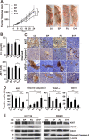
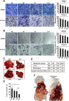
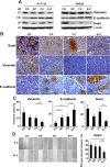
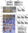
5-Fluorouracil (5-FU) is one of the most effective drugs for the treatment of colorectal cancer (CRC). However, there is an urgent need in reducing its systemic side effects and chemoresistance to make 5-FU-based chemotherapy more effective and less toxic in the treatment of CRC. Here, enalapril, a clinically widely used antihypertensive and anti-heart failure drug, has been verified as a chemosensitizer that extremely improves the sensitivity of CRC cells to 5-FU. Enalapril greatly augmented the cytotoxicity of 5-FU on the cell growth in both established and primary CRC cells. The combination of enalapril and 5-FU synergistically suppressed the cell migration and invasion in both 5-FU-sensitive and -resistant CRC cells in vitro, and inhibited angiogenesis, tumor growth, and metastasis of 5-FU-resistant CRC cells in vivo without increased systemic toxicity at concentrations that were ineffective as individual agents. Furthermore, combined treatment cooperatively inhibited NF-κB/STAT3 signaling pathway and subsequently reduced the expression levels of NF-κB/STAT3-regulated proteins (c-Myc, Cyclin D1, MMP-9, MMP-2, VEGF, Bcl-2, and XIAP) in vitro and in vivo. This study provides the first evidence that enalapril greatly sensitized CRC cells to 5-FU at clinically achievable concentrations without additional toxicity and the synergistic effect may be mainly by cooperatively suppressing proliferation, angiogenesis, and NF-κB/STAT3-regulated proteins.
Conflict of interest statement
The authors declare that they have no conflicts of interests.
Figures


Similar articles
-
Ursolic acid inhibits growth and metastasis of human colorectal cancer in an orthotopic nude mouse model by targeting multiple cell signaling pathways: chemosensitization with capecitabine.Clin Cancer Res. 2012 Sep 15;18(18):4942-53. doi: 10.1158/1078-0432.CCR-11-2805. Epub 2012 Jul 25. Clin Cancer Res. 2012. PMID: 22832932 Free PMC article.
-
Troxerutin (TXN) potentiated 5-Fluorouracil (5-Fu) treatment of human gastric cancer through suppressing STAT3/NF-κB and Bcl-2 signaling pathways.Biomed Pharmacother. 2017 Aug;92:95-107. doi: 10.1016/j.biopha.2017.04.059. Epub 2017 May 19. Biomed Pharmacother. 2017. PMID: 28531805
-
Esculetin enhances the inhibitory effect of 5-Fluorouracil on the proliferation, migration and epithelial-mesenchymal transition of colorectal cancer.Cancer Biomark. 2019;24(2):231-240. doi: 10.3233/CBM-181764. Cancer Biomark. 2019. PMID: 30689555
-
Current Perspectives on the Role of Nrf2 in 5-Fluorouracil Resistance in Colorectal Cancer.Anticancer Agents Med Chem. 2021;21(17):2297-2303. doi: 10.2174/1871520621666210129094354. Anticancer Agents Med Chem. 2021. PMID: 33511942 Review.
-
Beyond chemotherapy: Exploring 5-FU resistance and stemness in colorectal cancer.Eur J Pharmacol. 2025 Mar 15;991:177294. doi: 10.1016/j.ejphar.2025.177294. Epub 2025 Jan 23. Eur J Pharmacol. 2025. PMID: 39863147 Review.
Cited by
-
Iron Overloading Potentiates the Antitumor Activity of 5-Fluorouracil by Promoting Apoptosis and Ferroptosis in Colorectal Cancer Cells.Cell Biochem Biophys. 2024 Dec;82(4):3763-3780. doi: 10.1007/s12013-024-01463-x. Epub 2024 Aug 4. Cell Biochem Biophys. 2024. PMID: 39097854 Free PMC article.
-
In Vitro Drug Repurposing: Focus on Vasodilators.Cells. 2023 Feb 20;12(4):671. doi: 10.3390/cells12040671. Cells. 2023. PMID: 36831338 Free PMC article. Review.
-
Reposition of the anti-inflammatory drug diacerein in an in-vivo colorectal cancer model.Saudi Pharm J. 2022 Jan;30(1):72-90. doi: 10.1016/j.jsps.2021.12.009. Epub 2021 Dec 31. Saudi Pharm J. 2022. PMID: 35145347 Free PMC article.
-
Rutaecarpine suppresses the proliferation and metastasis of colon cancer cells by regulating the STAT3 signaling.J Cancer. 2022 Jan 1;13(3):847-857. doi: 10.7150/jca.66177. eCollection 2022. J Cancer. 2022. PMID: 35154453 Free PMC article.
-
Recent Updates on Mechanisms of Resistance to 5-Fluorouracil and Reversal Strategies in Colon Cancer Treatment.Biology (Basel). 2021 Aug 31;10(9):854. doi: 10.3390/biology10090854. Biology (Basel). 2021. PMID: 34571731 Free PMC article. Review.
References
-
- Siegel RL, et al. Colorectal cancer statistics, 2017. CA Cancer J. Clin. 2017;67:177–193. - PubMed
-
- Peeters M, et al. Randomized phase III study of panitumumab with fluorouracil, leucovorin, and irinotecan (FOLFIRI) compared with FOLFIRI alone as second-line treatment in patients with metastatic colorectal cancer. J. Clin. Oncol. 2010;28:4706–4713. - PubMed
-
- Vazquez C, et al. Prediction of severe toxicity in adult patients under treatment with 5-fluorouracil: a prospective cohort study. Anticancer Drugs. 2017;28:1039–1046. - PubMed
-
- Wilhelm M, et al. Prospective, multicenter study of 5-fluorouracil therapeutic drug monitoring in metastatic colorectal cancer treated in routine clinical practice. Clin. Colorectal Cancer. 2016;15(4):381–388. - PubMed
Publication types
MeSH terms
Substances
LinkOut - more resources
Full Text Sources
Medical
Research Materials
Miscellaneous

