. 2020 Jun 24;11(1):3276.
doi: 10.1038/s41467-020-16889-z.

Human GBP1 binds LPS to initiate assembly of a caspase-4 activating platform on cytosolic bacteria

Affiliations

Human GBP1 binds LPS to initiate assembly of a caspase-4 activating platform on cytosolic bacteria

José Carlos Santos et al. Nat Commun. .

Abstract

The human non-canonical inflammasome controls caspase-4 activation and gasdermin-D-dependent pyroptosis in response to cytosolic bacterial lipopolysaccharide (LPS). Since LPS binds and oligomerizes caspase-4, the pathway is thought to proceed without dedicated LPS sensors or an activation platform. Here we report that interferon-induced guanylate-binding proteins (GBPs) are required for non-canonical inflammasome activation by cytosolic Salmonella or upon cytosolic delivery of LPS. GBP1 associates with the surface of cytosolic Salmonella seconds after bacterial escape from their vacuole, initiating the recruitment of GBP2-4 to assemble a GBP coat. The GBP coat then promotes the recruitment of caspase-4 to the bacterial surface and caspase activation, in absence of bacteriolysis. Mechanistically, GBP1 binds LPS with high affinity through electrostatic interactions. Our findings indicate that in human epithelial cells GBP1 acts as a cytosolic LPS sensor and assembles a platform for caspase-4 recruitment and activation at LPS-containing membranes as the first step of non-canonical inflammasome signaling.


Conflict of interest statement

The authors declare no competing interests.

Figures

Fig. 1
Fig. 1. IFNγ priming is required for LPS-induced caspase-4 activation in human epithelial cells.
ac Intracellular bacterial fold-replication (a) and release of LDH (b, c) in naive or IFNγ-primed wild-type, CASP4–/– or GSDMD–/– HeLa cells, at 1 or 6-h post-infection (p.i.) with Salmonella. Cells in 96-well plates were infected for 30 min, washed and gentamicin was added to kill extracellular bacteria. At the indicated time points supernatant was collected to determine the release of LDH, and then cells were lysed and the number of viable intracellular bacteria was determined by counting colony forming units (CFUs). The bacterial fold-replication was calculated relative to 1 h p.i. d Percentage of CHQ-resistant cytosolic Salmonella in naive or IFNγ-primed HeLa at 1.5 h p.i. Cells were infected for 30 min as in (a) and then treated with gentamicin ± CHQ for an additional 1 h before cells were lysed and bacteria counted by CFUs. The percentage of cytosolic bacteria was calculated as the ratio of (CHQ + gentamicinresistant / gentamicinresistant). eg Release of LDH from naive or IFNγ-primed cells, 5 h after transfection with LPS (2.5 µg/50,000 cells) or 3–4 h after electroporation with LPS (300 ng/50,0000 cells). h Western blot analysis of full length (p43) and cleaved (p32) caspase-4 in the supernatants and cell lysates from naive or IFNγ-primed HaCaT cells, upon transfection with E. coli LPS LPS (2.5 µg/50,000 cells). i Streptavidin pull-down assay of the binding of biotin-conjugated LPS to endogenous caspase-4 from the lysates of naive or IFNγ-primed HBEC3-KT. Cells in 6-well plates were transfected with LPS-biotin (10 µg) or left untransfected, and biotinylated substrate was pulled down using equal amounts of streptavidin magnetic beads, which were then eluted in equal volumes of SDS-PAGE reducing sample buffer. Streptavidin-bound and -unbound fractions were analyzed by immunoblotting for caspase-4. Graphs show the mean ± SD, and data are pooled from two to six independent experiments performed in triplicate (ag) or representative of two (h, i) independent experiments. *** P < 0.001; ns, not significant; two-tailed t-test.
Fig. 2
Fig. 2. GBP1 is required for Salmonella- and LPS-induced caspase-4 activation to induce pyroptosis in epithelial cells.
a Immunoblots for GBP1, caspase-4 and GAPDH (loading control) in cell lysates from IFNγ-primed wild-type or GBP1–/– HeLa. b, c Release of LDH from naive or IFNγ-primed wild-type or GBP1–/– HeLa after Salmonella infection (b) or after 5 h transfection with E. coli LPS (2.5 µg / 50,000 cells) (c). d, e Immunoblots for full length (p43) and cleaved (p32) caspase-4 in combined supernatants and cell lysates from naive or IFNγ-primed wild-type and GBP1–/– HeLa, upon transfection with E. coli LPS for 5 h (d) or Salmonella infection (e). f Release of LDH from IFNγ-primed wild-type or GBP1–/– HeLa, 3 h after electroporation with LPS (300 ng/50,000 cells). g Release of LDH in IFNγ-primed HBEC3-KT or HaCaT cells treated with non-targeting control siRNA (NT) or with siRNAs targeting CASP4, GSDMD or GBP1, after E. coli LPS transfection. Cells were treated with siRNAs for 24 h and transfected with LPS (2.5 µg / 50,000 cells) for 5 h. Graphs show the mean ± SD, and data are pooled from two (c, f), three (g) or four (b) independent experiments performed in triplicate, or representative of three independent experiments (d, e). ***P < 0.001; ns, not significant; two-tailed t-test.
Fig. 3
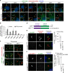
Fig. 3. GBP1 targets Salmonella and controls recruitment of GBP2-4.
a Fluorescence confocal microscopy of naive or IFNγ-primed HeLa expressing N-terminal tagged eGFP-GBP1-7 (green) and infected with Salmonella-dsRed (red) for 1 h. DNA was stained with Hoechst (blue). Representative confocal images are shown and scale bars correspond to 10 µm. b Percentage of intracellular Salmonella positive for eGFP-GBP1-7 in naive or IFNγ-primed HeLa, at 1 h p.i. At least 200–300 bacteria were counted per coverslip. c Fluorescence confocal microscopy of IFNγ-primed wild-type or GBP1–/– HeLa expressing eGFP-GBP2-4 (green) and infected with Salmonella-dsRed (red) for 1 h. Representative confocal images are shown, and scale bar corresponds to 10 µm. d Schematic representation of wild-type GBP1 and a ΔCaaX mutant. e, f Fluorescence confocal microscopy of naive GBP1–/– HeLa expressing HA-GBP1wt or HA-GBP1ΔCaaX (e) or co-expressing mCherry-GBP1wt and HA-GBP1wt or HA-GBP1ΔCaaX (f) and infected with Salmonella for 1 h. HA-tagged GBP1 was visualized by immunostaining with an anti-HA antibody. Representative confocal images are shown and scale bars correspond to 5 µm. The percentage of HA-GBP1 positive bacteria was quantified by counting around 100 bacteria per coverslip. Graphs show the mean ± SD, and data are pooled from two (b, e, f) independent experiments performed in duplicate or representative of two (a, c, e, f) independent experiments.
Fig. 4
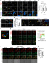
Fig. 4. GBP1 targets cytosolic Salmonella and is required for caspase-4 recruitment to the bacterial surface.
a Fluorescence confocal microscopy of IFNγ-primed wild-type HeLa co-expressing galectin-3-eGFP (green) and mCherry-GBP1-7 (red) and infected with Salmonella for 1 h. b Percentage of CHQ-resistant cytosolic Salmonella in naive or IFNγ-primed wild-type or GBP1–/– HeLa at 1.5 h p.i. Cells in triplicate wells were infected for 30 min and then treated with gentamicin ± CHQ for an additional 1 h before lysing the cells and determining CFUs. The percentage of cytosolic bacteria was calculated as the ratio of (CHQ + gentamicinresistant/gentamicinresistant). c Fluorescence confocal microscopy of naive or IFNγ-primed HeLa expressing caspase-4-eGFP (green) and infected with Salmonella-dsRed for 1 h. d. Percentage of caspase-4-eGFP positive Salmonella at 1 h p.i., quantified by counting 100–200 bacteria per coverslip. nd, not detected. e, f Fluorescence confocal microscopy of IFNγ-primed HeLa co-expressing galectin-3-mOrange (red) and caspase-4-eGFP (green) and infected with Salmonella for 1 h. g Time-lapse fluorescence confocal microscopy of IFNγ-primed HeLa expressing caspase-4-eGFP (green) and galectin-3-mOrange (red) and infected with Salmonella. h. Mean normalized fluorescence intensities of galectin-3-mOrange and caspase-4-eGFP over time. Fluorescence intensities were quantified in a region of interest as exemplified in the figure, containing an event of caspase-4 and galectin-3 recruitment to an individual bacterium. The relative intensity signals were aligned using the time point of onset of galectin-3 recruitment as zero and the mean and SD of six different events were plotted. i Time-lapse fluorescence confocal microscopy of IFNγ-primed HeLa co-expressing caspase-4-eGFP (green) and mCherry-GBP1 (red) and infected with Salmonella. Images were acquired every 60 s. DIC, differential interference contrast. j Percentage of caspase-4-eGFP positive Salmonella at 1 h p.i., in IFNγ-primed wild-type and GBP1–/– HeLa. 100–200 bacteria were counted per coverslip. Representative confocal images are shown and scale bars correspond to 1 µm (f), 5 µm (c, e, g) or 10 µm (a, i). Graphs show the mean ± SD, and data are pooled from two (b) or three (d, j) independent experiments performed in triplicate or representative from at least three independent experiments (a, c, ei). ***P < 0.001; ns, not significant, two-tailed t-test.
Fig. 5
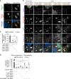
Fig. 5. GBP1/4 are sufficient to recruit caspase-4 to Salmonella and together with GBP3 activate the non-canonical inflammasome in naive human epithelial cells.
a Fluorescence confocal microscopy of naive and IFNγ-primed HeLa co-expressing caspase-4-eGFP (green) and mCherry-GBP1 (red) and infected with Salmonella for 1 h. DNA was stained with Hoechst (blue). Representative confocal images are shown and scale bar corresponds to 5 µm. b Fluorescence confocal microscopy of IFNγ-primed or naive HeLa cells co-expressing mCherry-GBP1 (red), Dox-inducible eGFP-GBP1, −2, −3 or −4 (green) and caspase-4-V5 (gray), and infected with Salmonella for 1 h. DNA was stained with Hoechst (blue). eGFP-GBPs were expressed by inducing cells with 1 µg/mL Dox for 16 h. Caspase-4-V5 was visualized by immunostaining with an anti-V5 antibody. Representative confocal images are shown and scale bar corresponds to 10 µm. c Percentage of caspase-4-V5 positive Salmonella at 1 h p.i., quantified out of the mCherry-GBP1-positive bacteria. At least 50 GBP1-positive bacteria were counted per coverslip. d Percentage of cell death in HeLa cells co-expressing constitutive mCherry-GBP1 and Dox-inducible eGFP or eGFP-GBP1, −2, −3 or −4. FLAG-GBP3 and HA-GBP4 were constitutively expressed together using a bicistronic plasmid. Cells were transfected with the indicated plasmids for 24 h. eGFP-GBPs were induced for 16 h with 1 µg/mL Dox, whereas eGFP was induced for 3 h. Cells were then transfected with E. coli-derived LPS (2.5 µg/50,000 cells) for 6 h and cell death values were normalized considering IFNγ-primed HeLa as 100% and naive cells co-expressing mCherry-GBP1 and eGFP as 0%. Graphs show the mean ± SD, and data are pooled from two independent experiments performed in duplicate (c), pooled from three independent experiments performed in triplicate (d) or are representative from two (b) or three (a) independent experiments. **P < 0.01; ***P < 0.001; one-way ANOVA.
Fig. 6
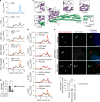
Fig. 6. LPS binds to GBP1 to induce formation of a high-molecular weight protein complex.
a Streptavidin pull-down assay for eGFP-GBP1-4 using biotin or biotin-conjugated LPS. HeLa cells stably expressing Dox-inducible eGFP-GBP1, −2, −3, or −4 were primed with IFNγ and 1 µg/mL Dox was added for 16 h. 1 million cells were lysed and incubated with 2 µg LPS-biotin or biotin, and the biotinylated substrates were pulled down using equal amounts of streptavidin magnetic beads, which were then eluted in equal volumes of SDS-PAGE reducing sample buffer. Streptavidin-bound and -unbound fractions were analyzed by western blot using an antibody against GFP. b SPR sensorgram of E. coli LPS (O111:B4) binding to human GBP1 immobilized on a CM5 chip surface. Sensorgram was obtained by using different LPS concentrations (47, 94, 188, 375, 750, and 1500 nM). Gray lines correspond to SPR data and orange lines to model fits using a two-state-reaction model. c Saturation curve of the titration of LPS on GBP1 immobilized on a CM5 chip. d Calculated dissociation constants (KD) for LPS binding to immobilized GBP1 (GBP1im) or GBP1 binding to immobilized E. coli LPS (LPSim). Dissociation constants for LPS-caspase-4 and LPS-caspase-11 were previously published by Shi et al.. e, f Size exclusion chromatograms of recombinant, LPS-free His-tagged GBP1 incubated with various LPS derivatives. Following purification, GBP1 (1 µM) was incubated on ice with LPS (2 µM) for 5 h before being subjected to size-exclusion analysis on a Superdex 200 10/30 GL column. Protein size was estimated using molecular weight standards. Curves were corrected by subtracting LPS-specific absorbance at 280 nm. Individual fractions were run on a 12% acrylamide gel and immunoblotted against His6 to confirm the presence of GBP1 in elution peaks (f). g GTPase activity analysis of recombinant GBP1. GBP1 (500 nM) was incubated with GTP (5 µM) with or without ultrapure LPS (5 µM) for 30 min before the reaction was stopped. Luminescence was normalized to a buffer-only control. Graphs show the mean ± SD, and data are representative from three (ad, g) or five (e, f) independent experiments performed with at least three independently expressed and purified batches of recombinant His-GBP1.
Fig. 7
Fig. 7. GBP1 is recruited to the bacterial surface and binds LPS through electrostatic interactions.
a Size exclusion chromatograms of recombinant His-tagged GBP1 incubated with E. coli LPS, or with E. coli LPS pre-treated with CaCl2 (5 mM), Polymyxin B (10 µg/mL) or with alkaline phosphatase. Curves were corrected by subtracting the respective LPS-specific absorbance at 280 nm. Black curves representing control condition were overlayed. b Release of LDH from IFNγ-primed HeLa 5 h after transfection with E. coli LPS, or after transfection with LPS previously treated with alkaline phosphatase. c 3D structure of human GBP1 (PDB 1f5n), highlighting five different negatively charged patches (A to E). Residues comprising patch E are only visible in PDB 6k1z. For each patch, the indicated residues were all mutated to alanines and analyzed for GBP1-LPS interaction by size exclusion chromatography. Purple indicates GTPase domain, green indicates helical domain. d Size exclusion chromatograms of different His-tagged GBP1 mutants incubated with E. coli LPS. Curves were corrected by subtracting LPS-specific absorbance at 280 nm. e Fluorescence confocal microscopy of naive HeLa expressing eGFP-GBP1wt, eGFP-GBP1KKK61-63AAA or eGFP-GBP1KK87-88AA and infected with Salmonella-dsRed for 1 h. DNA was stained with Hoechst (blue). Representative confocal images are shown and scale bar corresponds to 5 µm. f Percentage of eGFP-GBP1 positive Salmonella at 1 h p.i., as quantified by counting between 100–200 bacteria per coverslip. Graphs show the mean ± SD, and data are pooled from three independent experiments performed in duplicate (f), four independent experiments performed in triplicate (b), or are representative from three (a, d, e) independent experiments. ***P < 0.001; ns, not significant, two-tailed t-test.

Comment in

References

    1. Takeuchi O, Akira S. Pattern recognition receptors and inflammation. Cell. 2010;140:805–820. - PubMed
    1. Kayagaki N, et al. Caspase-11 cleaves gasdermin D for non-canonical inflammasome signalling. Nature. 2015;526:666–671. doi: 10.1038/nature15541. - DOI - PubMed
    1. Kayagaki N, et al. Noncanonical inflammasome activation by intracellular LPS independent of TLR4. Science. 2013;341:1246–1249. - PubMed
    1. Hagar JA, Powell DA, Aachoui Y, Ernst RK, Miao EA. Cytoplasmic LPS activates caspase-11: implications in TLR4-independent endotoxic shock. Science. 2013;341:1250–1253. - PMC - PubMed
    1. Shi J, et al. Inflammatory caspases are innate immune receptors for intracellular LPS. Nature. 2014;514:187–192. - PubMed

Publication types