

Lung-only melanoma: UV mutational signature supports origin from occult cutaneous primaries and argues against the concept of primary pulmonary melanoma
- PMID: 32581366
- PMCID: PMC8386291
- DOI: 10.1038/s41379-020-0594-0
Lung-only melanoma: UV mutational signature supports origin from occult cutaneous primaries and argues against the concept of primary pulmonary melanoma
Abstract
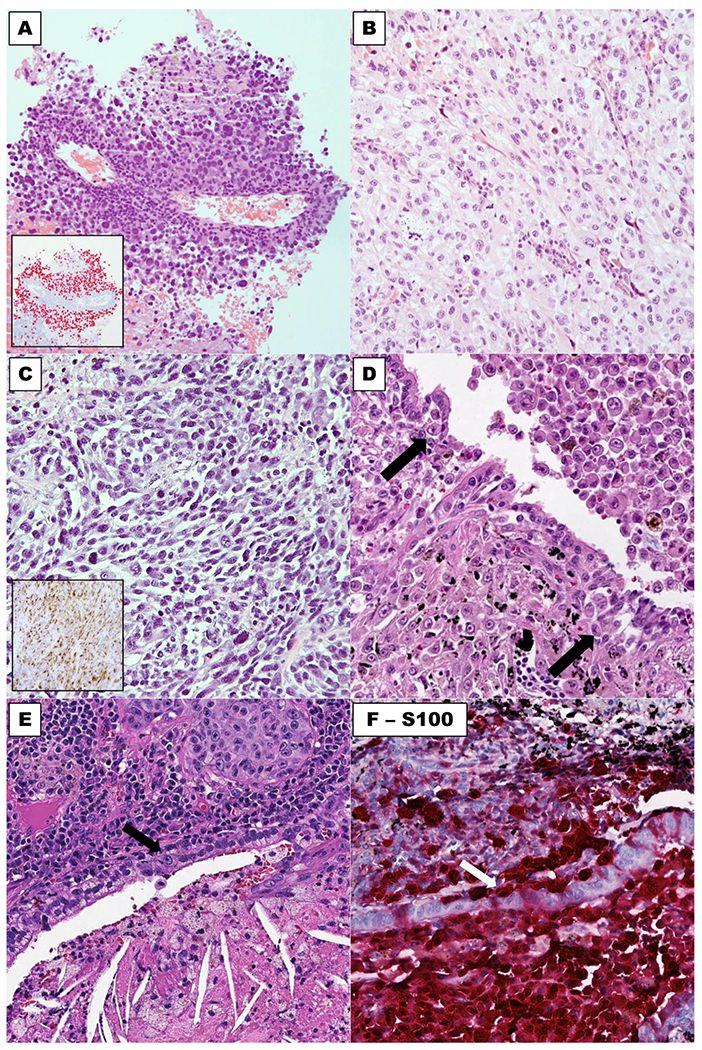
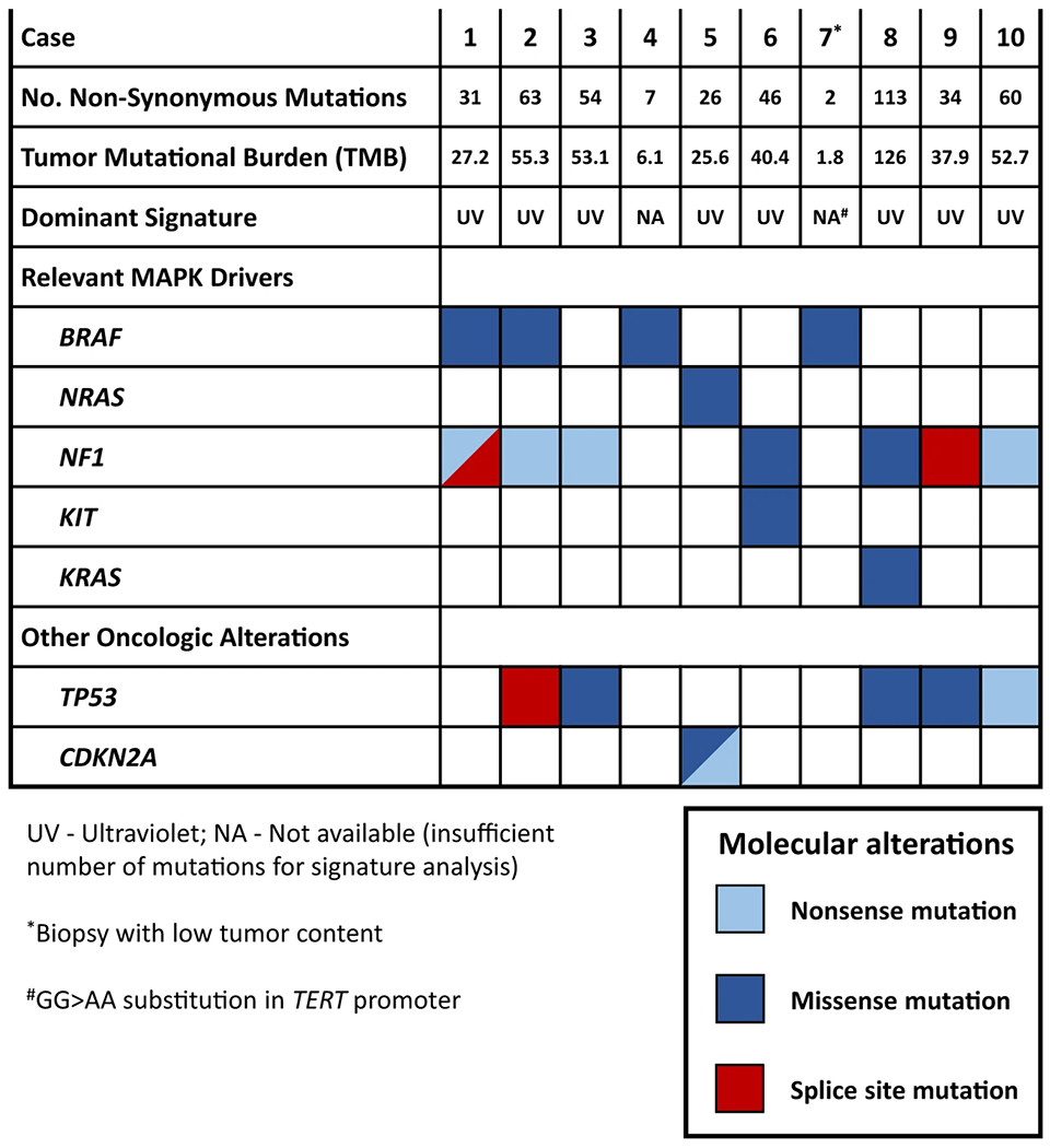
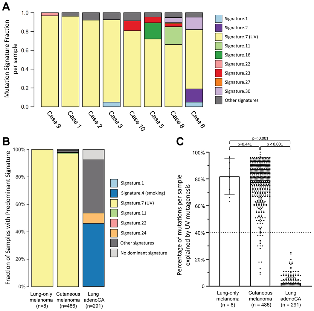
Primary pulmonary melanoma (PPM) is an entity recognized by the thoracic WHO classification. However, given the absence of native melanocytes in the lung and the known phenomenon of regression of cutaneous melanomas, the existence of PPM has remained controversial. Herein we investigate clinicopathologic and genomic features of lung-only melanomas with the goal to clarify their site of origin. We identified 10 melanomas involving exclusively lung with no current or previous cutaneous, uveal, or mucosal primaries. Four patients had solitary lesions with mean size of 5.1 cm (range 3.0-10.1 cm), meeting the criteria of PPM. Four patients had 2-3 lesions and 2 patients had >10 lesions. All cases underwent targeted next-generation sequencing interrogating up to 468 cancer genes, which revealed mean tumor mutation burden of 42.6 per megabase (range 1.8 to 126) and frequent mutations involving BRAF, NRAS, NF1, KIT, and KRAS - a genomic profile typical of UV-associated cutaneous melanoma. Mutational signature was assessable for eight cases harboring >20 mutations. This revealed that all evaluable cases harbored a dominant UV signature. In addition, one nonevaluable case harbored a GG > AA TERT promoter variant that is highly specific for UV-mutagenesis. As control groups, using the same methodology, a dominant UV signature was identified in 97% (470/486) of cutaneous melanomas, whereas no lung adenocarcinoma (n = 291) exhibited this signature. Notably, the clinical and pathologic features of solitary melanomas, especially those with large size and epithelioid morphology, closely mimicked primary lung carcinomas, highlighting a major potential for misdiagnosis. In conclusion, presence of a UV signature provides direct evidence that nearly all lung-only melanomas in this series, including solitary lesions meeting the strict criteria of PPM, represent metastases from occult cutaneous melanomas. This suggests that lung-only melanomas should be considered as likely metastatic even in the absence of a known primary melanoma elsewhere.
Conflict of interest statement
Figures

References
-
- Travis WD, Brambilla E, Burke AP, Marx A, Nicholson AG. WHO classification of tumours of the lung, pleura, thymus and heart, 4th ed. Lyon: International Agency for Research on Cancer, 2015. - PubMed
-
- Wilson RW, Moran CA. Primary melanoma of the lung: a clinicopathologic and immunohistochemical study of eight cases. Am J Surg Pathol. 1997;21:1196–202. - PubMed
-
- Ost D, Joseph C, Sogoloff H, Menezes G. Primary pulmonary melanoma: case report and literature review. Mayo Clin Proc. 1999;74:62–6. - PubMed
-
- Kundranda MN, Clark CT, Chaudhry AA, Chan V, Daw HA. Primary malignant melanoma of the lung: a case report and review of the literature. Clin Lung Cancer. 2006;7:279–81. - PubMed
Publication types
MeSH terms
Substances
Grants and funding
LinkOut - more resources
Full Text Sources
Medical
Research Materials
Miscellaneous
