

Gastric Adenomyosis: A Rare Cause of Pyloric Mass in Children
- PMID: 32581446
- PMCID: PMC7302453
- DOI: 10.4103/jiaps.JIAPS_44_19
Gastric Adenomyosis: A Rare Cause of Pyloric Mass in Children
Abstract
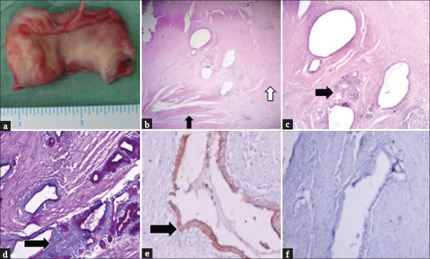
Adenomyoma of the stomach is a benign tumor with a very low incidence. Clinical presentation and imaging modalities are usually nonspecific and variable. A rare case of gastric adenomyoma in a 12-year-old child is being reported who presented with gastric outlet obstruction. The diagnosis could only be established after an excision biopsy performed after multiple diagnostic modalities failed to clinch the diagnosis. The case is being reported in view of the rarity of this entity in the pediatric age group as a cause of gastric outlet obstruction.
Keywords: Adenomyoma; gastric outlet obstruction; myoepithelial hamartoma.
Copyright: © 2020 Journal of Indian Association of Pediatric Surgeons.
Conflict of interest statement
There are no conflicts of interest.
Figures

Similar articles
-
Benign pyloric adenomyoma presented as gastric outlet obstruction: a case report and review of the literature.J Med Case Rep. 2024 Aug 24;18(1):397. doi: 10.1186/s13256-024-04741-7. J Med Case Rep. 2024. PMID: 39180137 Free PMC article. Review.
-
[Infantile hypertrofic pyloric stenosis or gastric adenomyoma? Differential diagnosis of gastric outlet obstruction in children].Cir Pediatr. 2015 Jul 20;28(3):153-155. Cir Pediatr. 2015. PMID: 27775311 Spanish.
-
Radiological findings of gastric adenomyoma in a neonate presenting with gastric outlet obstruction.Pediatr Radiol. 2013 Mar;43(5):628-30. doi: 10.1007/s00247-012-2521-0. Epub 2012 Oct 7. Pediatr Radiol. 2013. PMID: 23052729
-
Adenomyoma of the stomach mimicking infantile hypertrophic pyloric stenosis.J Pediatr Surg. 2007 Nov;42(11):E11-2. doi: 10.1016/j.jpedsurg.2007.07.050. J Pediatr Surg. 2007. PMID: 18022419
-
Report of three gastric tumors in children.J Pediatr Surg. 1994 Sep;29(9):1202-4. doi: 10.1016/0022-3468(94)90800-1. J Pediatr Surg. 1994. PMID: 7807345 Review.
Cited by
-
Gastric Adenomyoma with Heterotopic Pancreatic Tissue.J Indian Assoc Pediatr Surg. 2022 Mar-Apr;27(2):273-274. doi: 10.4103/jiaps.JIAPS_102_20. Epub 2022 Mar 1. J Indian Assoc Pediatr Surg. 2022. PMID: 35937125 Free PMC article. No abstract available.
-
Asymptomatic gastric adenomyoma and heterotopic pancreas in a patient with pancreatic cancer: A case report and review of the literature.World J Clin Cases. 2021 Sep 26;9(27):8147-8156. doi: 10.12998/wjcc.v9.i27.8147. World J Clin Cases. 2021. PMID: 34621874 Free PMC article.
-
Nonpolypous Hamartomas of the Gastrointestinal Tract: An Updated Review on Classification, Denominations, and Clinical Management.J Oncol. 2022 May 9;2022:6983460. doi: 10.1155/2022/6983460. eCollection 2022. J Oncol. 2022. PMID: 35586207 Free PMC article. Review.
-
Benign pyloric adenomyoma presented as gastric outlet obstruction: a case report and review of the literature.J Med Case Rep. 2024 Aug 24;18(1):397. doi: 10.1186/s13256-024-04741-7. J Med Case Rep. 2024. PMID: 39180137 Free PMC article. Review.
References
Publication types
LinkOut - more resources
Full Text Sources