

Evaluation of Left Main Coronary Artery Using Optical Frequency Domain Imaging and Its Pitfalls
- PMID: 32581660
- PMCID: PMC7306070
- DOI: 10.1155/2020/4817239
Evaluation of Left Main Coronary Artery Using Optical Frequency Domain Imaging and Its Pitfalls
Abstract
Objectives: We aimed to assess the quality of optical frequency domain imaging (OFDI) of the left main (LM) arterial wall and describe and analyse potential artefacts in this setting.
Background: OFDI is increasingly used to assess ambiguous lesions and optimize LM percutaneous coronary intervention. However, its ability to provide artefact-free high-quality images of coronary ostia and large segments such as the LM remains uncertain.
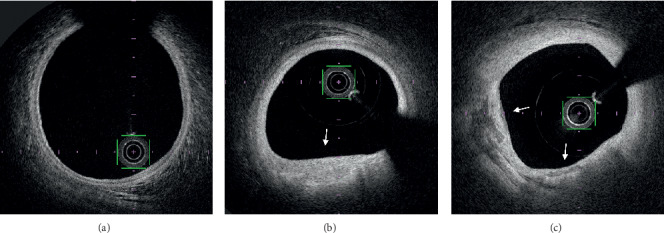
Methods: We included 42 consecutive patients who underwent OFDI, including LM imaging. Each OFDI frame was subdivided into four quadrants and analysed. The number of quadrants with artifacts was calculated within the proximal, mid, and distal LM and the first 5 mm of the left anterior descending artery (LAD) and/or left circumflex artery (LCX).
Results: The quadrants analysis showed an overall artifact rate of 8.9%, mostly out-of-field (45.1%) or residual blood (44.7%) artefacts. Most artifacts were located in the proximal LM (18.6%) with a stepwise reduction of artifact rates towards distal segments (mid LM 5.8%; distal LM 3.6%, ostial LAD 2.6%, and ostial LCX 0%; p < 0.001). While 20 (48.8%) patients had angiographically visible plaques, OFDI showed plaques in 32 patients (76.2%; p=0.007).
Conclusion: OFDI can accurately evaluate the LM and detect and assess angiographically unvisualized atherosclerotic plaques providing accurate assessment of >90% of the quadrants of the LM and the ostia of its bifurcation branches. However, artifacts mainly located in the proximal LM and decreasing distally in a stepwise fashion should be considered in the interpretation of OFDI in this setting.
Copyright © 2020 Vincent Roule et al.
Conflict of interest statement
The authors have no conflicts of interest to declare.
Figures


References
-
- Giannoglou G. D., Antoniadis A. P., Chatzizisis Y. S., Damvopoulou E., Parcharidis G. E., Louridas G. E. Prevalence of narrowing >or = 50% of the left main coronary artery among 17,300 patients having coronary angiography. The American Journal of Cardiology. 2006;98(9):1202–1205. - PubMed
-
- Isner J. M., Kishel J., Kent K. M., Ronan J. A., Jr., Ross A. M., Roberts W. C. Accuracy of angiographic determination of left main coronary arterial narrowing. Angiographic–histologic correlative analysis in 28 patients. Circulation. 1981;63(5):1056–1064. doi: 10.1161/01.cir.63.5.1056. - DOI - PubMed
MeSH terms
LinkOut - more resources
Full Text Sources
Medical
