Review
. 2020 Jun 9:14:571.
doi: 10.3389/fnins.2020.00571. eCollection 2020.

Gene Deregulation and Underlying Mechanisms in Spinocerebellar Ataxias With Polyglutamine Expansion

Affiliations
Review

Gene Deregulation and Underlying Mechanisms in Spinocerebellar Ataxias With Polyglutamine Expansion

Anna Niewiadomska-Cimicka et al. Front Neurosci. .

Abstract

Polyglutamine spinocerebellar ataxias (polyQ SCAs) include SCA1, SCA2, SCA3, SCA6, SCA7, and SCA17 and constitute a group of adult onset neurodegenerative disorders caused by the expansion of a CAG repeat sequence located within the coding region of specific genes, which translates into polyglutamine tract in the corresponding proteins. PolyQ SCAs are characterized by degeneration of the cerebellum and its associated structures and lead to progressive ataxia and other diverse symptoms. In recent years, gene and epigenetic deregulations have been shown to play a critical role in the pathogenesis of polyQ SCAs. Here, we provide an overview of the functions of wild type and pathogenic polyQ SCA proteins in gene regulation, describe the extent and nature of gene expression changes and their pathological consequences in diseases, and discuss potential avenues to further investigate converging and distinct disease pathways and to develop therapeutic strategies.

Keywords: Purkinje cells; SCA; epigenetic; polyglutamine; spinocerebellar ataxia; transcriptional dysregulation.


Figures

FIGURE 1
FIGURE 1
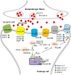
Schematic of Purkinje cell enriched gene deregulations in polyQ SCAs affecting glutamatergic synapse. Deregulations of gene expression in Purkinje cells result in alterations of the protein level of ion channels, glutamate transporters, and receptors together with downstream effectors. Production of secondary messengers and calcium homeostasis are impaired, which leads to alteration of long-term depression and pacemaker activities.
FIGURE 2
FIGURE 2
Wild type and pathological functions of polyQ SCA proteins in gene expression. Wild type SCA proteins participate to various regulatory steps of gene expression, such as binding to DNA sequences (α1ACT and TBP), modulating repression or activation activities of transcription factors (TF) (ATXN1, ATXN3, ATXN7, and TBP), modifying epigenetic marks on promoters (ATXN7/SAGA and ATXN3/HDAC3/SMRT/NCOR complexes), assembling pre-initiation complex (PIC) (TBP/TFIID) that includes general transcription factors (GTFs), deubiquitinating substrates (ATXN3 and ATXN7/SAGA) and participating to RNA metabolism (ATXN1 and ATXN2). Due to abnormal interactions with their native partners, mutant SCA proteins alter (1) the activation or repressor activities of TFs, (2) the epigenetic marks of histones involved in chromatin opening, (3) the formation of PIC recruiting RNAPII on gene promoters, and (4) the transcription elongation mediated by phosphorylated RNAPII (p-RNAPII) and by histone modification on the gene body. Mutant SCA proteins also alter (5) the post-translation modification (PTM) of nuclear proteins, (6) RNA splicing and (7) other RNA processing such as translation. In addition, (8) mutant SCA proteins form soluble oligomers and insoluble aggregates that sequester native protein partners and proteins involved in nuclear proteostasis, accounting for perturbation of gene expression. ub, ubiquitin; ac, acetyl; RBP, ribosome binding proteins.

References

    1. Abou-Sleymane G., Chalmel F., Helmlinger D., Lardenois A., Thibault C., Weber C., et al. (2006). Polyglutamine expansion causes neurodegeneration by altering the neuronal differentiation program. Hum. Mol. Genet. 15 691–703. 10.1093/hmg/ddi483 - DOI - PubMed
    1. Achour M., Le Gras S., Keime C., Parmentier F., Lejeune F. X., Boutillier A. L., et al. (2015). Neuronal identity genes regulated by super-enhancers are preferentially down-regulated in the striatum of Huntington’s disease mice. Hum. Mol. Genet. 24 3481–3496. 10.1093/hmg/ddv099 - DOI - PubMed
    1. Aikawa T., Mogushi K., Iijima-Tsutsui K., Ishikawa K., Sakurai M., Tanaka H., et al. (2015). Loss of MyD88 alters neuroinflammatory response and attenuates early Purkinje cell loss in a spinocerebellar ataxia type 6 mouse model. Hum. Mol. Genet. 24 4780–4791. 10.1093/hmg/ddv202 - DOI - PMC - PubMed
    1. Alves-Cruzeiro J. M., Mendonca L., Pereira de Almeida L., Nobrega C. (2016). Motor dysfunctions and neuropathology in mouse models of spinocerebellar ataxia type 2: a comprehensive review. Front. Neurosci. 10:572. 10.3389/fnins.2016.00572 - DOI - PMC - PubMed
    1. Araujo J., Breuer P., Dieringer S., Krauss S., Dorn S., Zimmermann K., et al. (2011). FOXO4-dependent upregulation of superoxide dismutase-2 in response to oxidative stress is impaired in spinocerebellar ataxia type 3. Hum. Mol. Genet. 20 2928–2941. 10.1093/hmg/ddr197 - DOI - PubMed