. 2020 Jun 11:14:644.
doi: 10.3389/fnins.2020.00644. eCollection 2020.

Loss of Wwox Perturbs Neuronal Migration and Impairs Early Cortical Development

Affiliations

Loss of Wwox Perturbs Neuronal Migration and Impairs Early Cortical Development

Michele Iacomino et al. Front Neurosci. .

Abstract

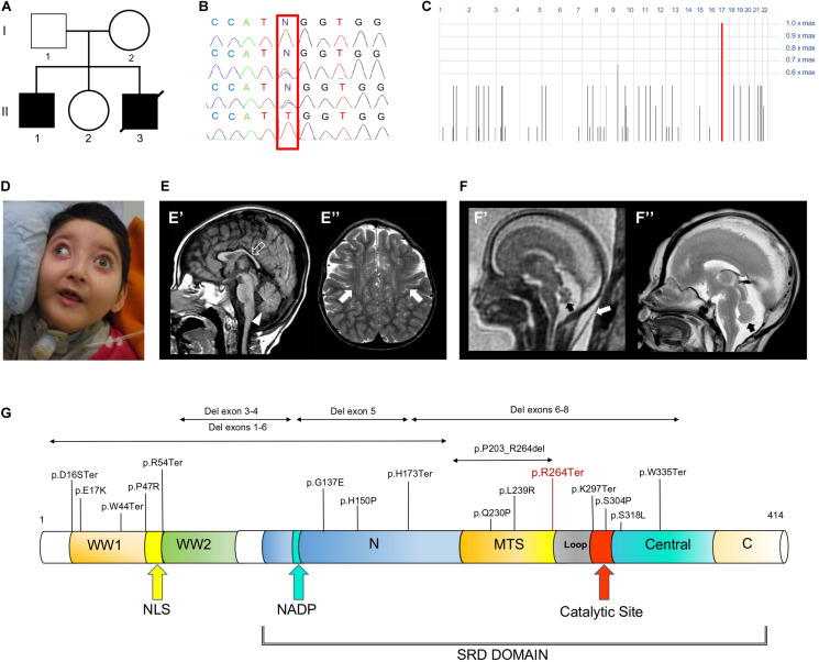
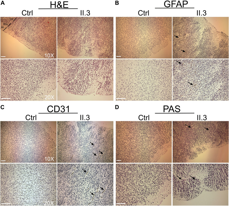
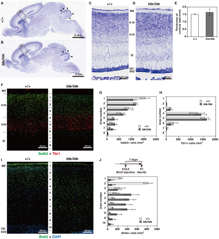
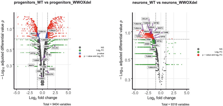
Mutations in the WWOX gene cause a broad range of ultra-rare neurodevelopmental and brain degenerative disorders, associated with a high likelihood of premature death in animal models as well as in humans. The encoded Wwox protein is a WW domain-containing oxidoreductase that participates in crucial biological processes including tumor suppression, cell growth/differentiation and regulation of steroid metabolism, while its role in neural development is less understood. We analyzed the exomes of a family affected with multiple pre- and postnatal anomalies, including cerebellar vermis hypoplasia, severe neurodevelopmental impairment and refractory epilepsy, and identified a segregating homozygous WWOX mutation leading to a premature stop codon. Abnormal cerebral cortex development due to a defective architecture of granular and molecular cell layers was found in the developing brain of a WWOX-deficient human fetus from this family. A similar disorganization of cortical layers was identified in lde/lde rats (carrying a homozygous truncating mutation which disrupts the active Wwox C-terminal domain) investigated at perinatal stages. Transcriptomic analyses of Wwox-depleted human neural progenitor cells showed an impaired expression of a number of neuronal migration-related genes encoding for tubulins, kinesins and associated proteins. These findings indicate that loss of Wwox may affect different cytoskeleton components and alter prenatal cortical development, highlighting a regulatory role of the WWOX gene in migrating neurons across different species.

Keywords: WOREE syndrome; WWOX; animal model; cytoskeleton; developing brain; neuropathology.


Figures

FIGURE 1
FIGURE 1
Family tree, genetic studies, WOREE-associated clinico-radiological features, Wwox protein and WOREE-associated mutations. (A) Pedigree from Family. (B) Electropherograms of carrier parents and index case (II.1) with the c.790C > T homozygous variant introducing a p.Arg264Ter stop codon. (C) Homozygosity mapping reveal a homozygous bloc in the WWOX gene 16q23.2 region. (D) Distinctive craniofacial features in WOREE syndrome due to homozygous p.Arg264Ter variant, including round hypotonic face and short neck. (E) Brain MRI of Patient II.1 performed at 2.4 years of age. (E’) Sagittal T1-weighted image reveals hypoplasia of the corpus callosum (empty arrow) and inferior cerebellar vermis (arrowhead). (E”) Axial T2-weighted images demonstrates mild atrophy of the frontal lobes with associated bilateral white matter hyperintensity (arrows). (F) Fetal MRI (F’) and high-resolution post-mortem MRI (F”) studies of Patient II.3 performed at 21 gestational weeks demonstrate mild hypoplasia of the cerebellar vermis (black arrows). Note the slightly increased thickness of nuchal subcutaneous tissues on fetal MRI (white arrow). The laminar organization of the cerebral hemispheres and cortical gyration are appropriate for the gestational age (not shown). (G) Wwox protein structure and domains and WOREE-associated mutations identified so far.
FIGURE 2
FIGURE 2
Histopathological studies of WWOX-deficient human brain. (A) H&E staining shows an incorrect migration of the external granular layer within the molecular layer in patient II.3 (right panel) compared to a normal fetus (left panel) at the same gestational age. Images at 10X and 20X magnification (molecular layer: ML, external granular layer: EGL, cortical gray: CG). Scale bar: 50 μm. (B) Histological staining with GFAP 10× (upper panel) and 20× (lower panel) in which a disorganization of the irregularly distributed trajectories is observed (black arrows). Scale bars: 50 μm. (C) Histological staining with CD-31 at 10× (upper panel) and 20× (lower panel), in which thinning of the vessels is observed in the context of the external granular layer (black arrows). (D) Histological staining with Shift Reactive Periodic Acid (PAS) at 10× (upper panel) and 20× (lower panel) show that the vascular structures of the cortex are irregularly distributed and irregularly branched (black arrows). Scale bars 50 μm.
FIGURE 3
FIGURE 3
Cortical layer formation and neural migration in lde/lde rat brain compared to normal one. (A–D) Sagittal brain sections at P1 rats were subjected to Nissl staining. Squares as labeled C and D in panels (A,B) indicated identical areas as shown in panels (C,D), respectively. Arrowheads indicate delayed foliation in lde/lde cerebellum. (E) The thicknesses of cerebral wall (MZ to VZ/SVZ) is shown as mean ± S.D. from three independent experiments. Student’s t-test is used to determine statistical significance (p < 0.05). (F) Sections are co-immunostained with anti-Satb2 (green) anti-Tbr1 (red). (I) Distribution of migrating neuronal cells labeled with BrdU (green) in cerebral cortex. Nuclei are counterstained with DAPI (blue). Representative images from three independent experiments are shown. The cell densities of Satb2 (G), Tbr1 (H), and BrdU-positive cells (J) in each area are plotted as histograms. Values are shown as the mean ± SD. The asterisk indicates a significant difference between +/+ and lde/lde in the same area by Student’s t-test (p < 0.05, ∗∗p < 0.01). Experimental scheme to label migrating cells by BrdU is illustrated in panel (J). MZ, marginal zone; IZ, intermediate zone; VZ, ventricular zone: SVZ, subventricular zone.
FIGURE 4
FIGURE 4
Wwox-depleted neurons show reduced expression of different neural migration-related genes. The differential gene expression profiles in control hNPC vs. WWOX-depleted hNPC and control neurons vs. WWOX-depleted neurons represented in Volcano Plot. P – significance was calculated according to NOISeq-sim algorithm.

References

    1. Abdel-Salam G., Thoenes M., Afifi H. H., Korber F., Swan D., Bolz H. J. (2014). The supposed tumor suppressor gene WWOX is mutated in an early lethal microcephaly syndrome with epilepsy, growth retardation and retinal degeneration. Orphanet J. Rare Dis. 9:12. 10.1186/1750-1172-9-12 - DOI - PMC - PubMed
    1. Aldaz C. M., Ferguson B. W., Abba M. C. (2014). WWOX at the crossroads of cancer, metabolic syndrome related traits and CNS pathologies. Biochim. Biophys. Acta. 1846 188–200. 10.1016/j.bbcan.2014.06.001 - DOI - PMC - PubMed
    1. Aqeilan R. I., Hagan J. P., de Bruin A., Rawahneh M., Salah Z., Gaudio E., et al. (2009). Targeted ablation of the WW domain-containing oxidoreductase tumor suppressor leads to impaired steroidogenesis. Endocrinology. 150 1530–1535. 10.1210/en.2008-1087 - DOI - PMC - PubMed
    1. Aqeilan R. I., Trapasso F., Hussain S., Costinean S., Marshall D., Pekarsky Y., et al. (2007). Targeted deletion of Wwox reveals a tumor suppressor function. PNAS 104 3949–3954. 10.1073/pnas.0609783104 - DOI - PMC - PubMed
    1. Arisio R., Bonissone M., Piccoli E., Panzica G. (2002). Central nervous system microangioarchitecture in the human foetus. Adv. Clin. Path 6 125–129. - PubMed