

GSK-3β Contributes to Parkinsonian Dopaminergic Neuron Death: Evidence From Conditional Knockout Mice and Tideglusib
- PMID: 32581704
- PMCID: PMC7283909
- DOI: 10.3389/fnmol.2020.00081
GSK-3β Contributes to Parkinsonian Dopaminergic Neuron Death: Evidence From Conditional Knockout Mice and Tideglusib
Abstract
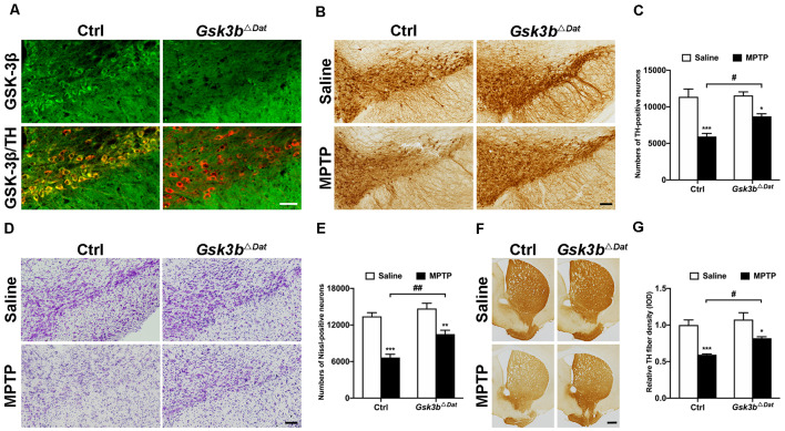
Glycogen synthase kinase-3 (GSK-3) dysregulation has been implicated in nigral dopaminergic neurodegeneration, one of the main pathological features of Parkinson's disease (PD). The two isoforms, GSK-3α and GSK-3β, have both been suggested to play a detrimental role in neuronal death. To date, several studies have focused on the role of GSK-3β on PD pathogenesis, while the role of GSK-3α has been largely overlooked. Here, we report in situ observations that both GSK-3α and GSK-3β are dephosphorylated at a negatively acting regulatory serine, indicating kinase activation, selectively in nigral dopaminergic neurons following exposure of mice to 1-methyl-4-pheny-1,2,3,6-tetrahydropyridine (MPTP). To identify whether GSK-3α and GSK-3β display functional redundancy in regulating parkinsonian dopaminergic cell death, we analysed dopaminergic neuron-specific Gsk3a null (Gsk3a ΔDat ) and Gsk3b null (Gsk3b ΔDat ) mice, respectively. We found that Gsk3b ΔDat , but not Gsk3a ΔDat , showed significant resistance to MPTP insult, revealing non-redundancy of GSK-3α and GSK-3β in PD pathogenesis. In addition, we tested the neuroprotective effect of tideglusib, the most clinically advanced inhibitor of GSK-3, in the MPTP model of PD. Administration of higher doses (200 mg/kg and 500 mg/kg) of tideglusib exhibited significant neuroprotection, whereas 50 mg/kg tideglusib failed to prevent dopaminergic neurodegeneration from MPTP toxicity. Administration of 200 mg/kg tideglusib improved motor symptoms of MPTP-treated mice. Together, these data demonstrate GSK-3β and not GSK-3α is critical for parkinsonian neurodegeneration. Our data support the view that GSK-3β acts as a potential therapeutic target in PD and tideglusib would be a candidate drug for PD neuroprotective therapy.
Keywords: GSK-3α; GSK-3β; MPTP; Parkinson’s disease; neuroprotection; tideglusib.
Copyright © 2020 Li, Ma, Chen, Hu, Li, Zhang, Su, Woodgett, Li and Huang.
Figures


Similar articles
-
GSK-3 mediates nuclear translocation of p62/SQSTM1 in MPTP-induced mouse model of Parkinson's disease.Neurosci Lett. 2021 Oct 15;763:136177. doi: 10.1016/j.neulet.2021.136177. Epub 2021 Aug 13. Neurosci Lett. 2021. PMID: 34400288
-
Multitarget intervention of Fasudil in the neuroprotection of dopaminergic neurons in MPTP-mouse model of Parkinson's disease.J Neurol Sci. 2015;353(1-2):28-37. doi: 10.1016/j.jns.2015.03.022. Epub 2015 Mar 20. J Neurol Sci. 2015. PMID: 25908255
-
L-F001, a Multifunction ROCK Inhibitor Prevents 6-OHDA Induced Cell Death Through Activating Akt/GSK-3beta and Nrf2/HO-1 Signaling Pathway in PC12 Cells and Attenuates MPTP-Induced Dopamine Neuron Toxicity in Mice.Neurochem Res. 2017 Feb;42(2):615-624. doi: 10.1007/s11064-016-2117-4. Epub 2017 Jan 11. Neurochem Res. 2017. PMID: 28078613
-
Association of glycogen synthase kinase-3β with Parkinson's disease (review).Mol Med Rep. 2014 Jun;9(6):2043-50. doi: 10.3892/mmr.2014.2080. Epub 2014 Mar 28. Mol Med Rep. 2014. PMID: 24681994 Free PMC article. Review.
-
Therapeutic Potential Effect of Glycogen Synthase Kinase 3 Beta (GSK-3β) Inhibitors in Parkinson Disease: Exploring an Overlooked Avenue.Mol Neurobiol. 2024 Sep;61(9):7092-7108. doi: 10.1007/s12035-024-04003-z. Epub 2024 Feb 17. Mol Neurobiol. 2024. PMID: 38367137 Free PMC article. Review.
Cited by
-
Glycogen Synthase Kinase 3: Ion Channels, Plasticity, and Diseases.Int J Mol Sci. 2022 Apr 16;23(8):4413. doi: 10.3390/ijms23084413. Int J Mol Sci. 2022. PMID: 35457230 Free PMC article. Review.
-
Molecular Determinants of A9 Dopaminergic Neurons.Neuromolecular Med. 2025 May 21;27(1):43. doi: 10.1007/s12017-025-08861-1. Neuromolecular Med. 2025. PMID: 40397062 Review.
-
TFE3-Mediated Autophagy is Involved in Dopaminergic Neurodegeneration in Parkinson's Disease.Front Cell Dev Biol. 2021 Nov 29;9:761773. doi: 10.3389/fcell.2021.761773. eCollection 2021. Front Cell Dev Biol. 2021. PMID: 34912803 Free PMC article.
-
Evaluation of Potential Neuroprotective Effects of Vanillin Against MPP+/MPTP-Induced Dysregulation of Dopaminergic Regulatory Mechanisms in SH-SY5Y Cells and a Mouse Model of Parkinson's Disease.Mol Neurobiol. 2023 Aug;60(8):4693-4715. doi: 10.1007/s12035-023-03358-z. Epub 2023 May 5. Mol Neurobiol. 2023. PMID: 37145378
-
The Common Denominators of Parkinson's Disease Pathogenesis and Methamphetamine Abuse.Curr Neuropharmacol. 2024;22(13):2113-2156. doi: 10.2174/1570159X21666230907151226. Curr Neuropharmacol. 2024. PMID: 37691228 Free PMC article. Review.
References
-
- Anagnostou E., Bennett T. A., Thorpe K., Nicolson R. (2018). 5.16 A phase 2 randomized, placebo-controlled trial of tideglusib, an orally administered GSK-3 β inhibitor, in the treatment of adolescents with ASD. J. Am. Acad. Child Adolesc. Psychiatry 57:S232 10.1016/j.jaac.2018.09.311 - DOI
-
- Avrahami L., Farfara D., Shaham-Kol M., Vassar R., Frenkel D., Eldar-Finkelman H. (2013). Inhibition of glycogen synthase kinase-3 ameliorates β-amyloid pathology and restores lysosomal acidification and mammalian target of rapamycin activity in the Alzheimer disease mouse model: in vivo and in vitro studies. J. Biol. Chem. 288, 1295–1306. 10.1074/jbc.m112.409250 - DOI - PMC - PubMed
LinkOut - more resources
Full Text Sources
Molecular Biology Databases
Miscellaneous