Review
. 2020 Jun 9:14:162.
doi: 10.3389/fncel.2020.00162. eCollection 2020.

Sialylation and Galectin-3 in Microglia-Mediated Neuroinflammation and Neurodegeneration

Affiliations
Review

Sialylation and Galectin-3 in Microglia-Mediated Neuroinflammation and Neurodegeneration

Mar Puigdellívol et al. Front Cell Neurosci. .

Abstract

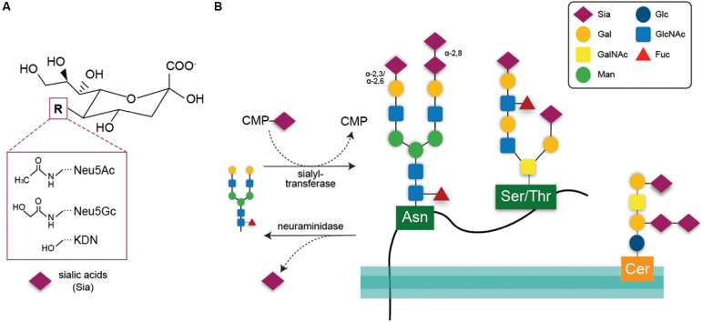
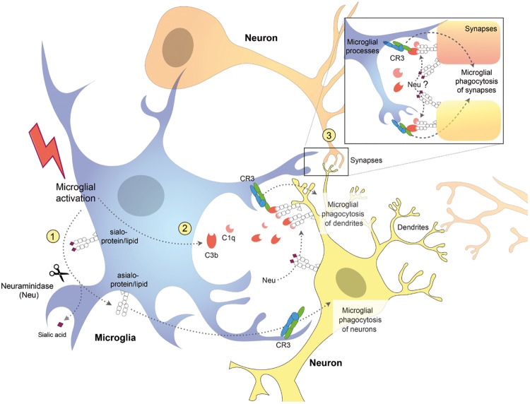
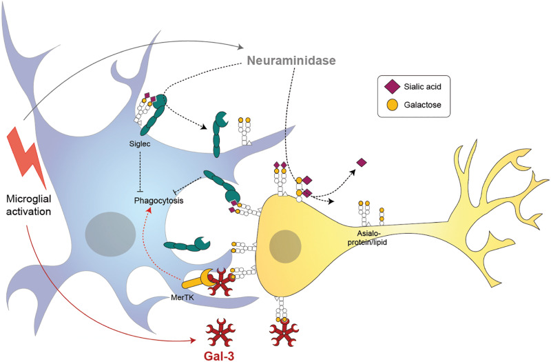
Microglia are brain macrophages that mediate neuroinflammation and contribute to and protect against neurodegeneration. The terminal sugar residue of all glycoproteins and glycolipids on the surface of mammalian cells is normally sialic acid, and addition of this negatively charged residue is known as "sialylation," whereas removal by sialidases is known as "desialylation." High sialylation of the neuronal cell surface inhibits microglial phagocytosis of such neurons, via: (i) activating sialic acid receptors (Siglecs) on microglia that inhibit phagocytosis and (ii) inhibiting binding of opsonins C1q, C3, and galectin-3. Microglial sialylation inhibits inflammatory activation of microglia via: (i) activating Siglec receptors CD22 and CD33 on microglia that inhibit phagocytosis and (ii) inhibiting Toll-like receptor 4 (TLR4), complement receptor 3 (CR3), and other microglial receptors. When activated, microglia release a sialidase activity that desialylates both microglia and neurons, activating the microglia and rendering the neurons susceptible to phagocytosis. Activated microglia also release galectin-3 (Gal-3), which: (i) further activates microglia via binding to TLR4 and TREM2, (ii) binds to desialylated neurons opsonizing them for phagocytosis via Mer tyrosine kinase, and (iii) promotes Aβ aggregation and toxicity in vivo. Gal-3 and desialylation may increase in a variety of brain pathologies. Thus, Gal-3 and sialidases are potential treatment targets to prevent neuroinflammation and neurodegeneration.

Keywords: aging; desialylation; galectin-3; microglia; neurodegeneration; phagocytosis; sialic acid.


Figures

FIGURE 1
FIGURE 1
(A) Chemical structure of the three core sialic acids, N-acetylneuraminic acid (Neu5Ac), N-glycolylneuraminic acid (Neu5Gc), and deaminoneuraminic acid (KDN). These core structures may be further modified, e.g., by sulfation or methylation. (B) Sialic acids are transferred onto acceptor glycans via sialyl-transferase enzymes in the Golgi that use the activated cytidine-5’-monophosphate-sialic acid (CMP-Sia) as a donor molecule. Sialyl residues may be added terminally to galactose residues in α-2,3 or α-2,6 linkage or to sialic acid residues in α-2,8 linkage. Such glycan chains may be attached to glycoproteins via asparagine residue (N-glycan) or to serine or threonine residues (O-glycan). Glycosylation of lipids is exemplified here by a ganglioside composed of ceramide (Cer) to an oligosaccharide chain. Sialic acids may be released via hydrolytic enzymes termed neuraminidases. Gal, galactose; GalNAc, N-acetylgalactosamine; Man, mannose; Glc, glucose; GlcNAc, N-acetylglucosamine; Fuc, fucose.
FIGURE 2
FIGURE 2
Schematic diagram showing potential mechanisms for complement receptor 3 (CR3)-dependent microglial phagocytosis of neurons, dendrites, and synapses. Activated microglia (1) desialylate their surface via neuraminidase (Neu) which stimulates microglial phagocytosis of neurons via CR3 (Allendorf et al., 2020b) and (2) release complement proteins C1q and C3b, which opsonize desialylated neuronal dendrites and (3) synapses, stimulating their phagocytosis via microglial CR3 (Linnartz et al., 2012). Neuraminidase released from microglia or onto the surface of neurons, desialylates the neuronal surface, and promotes binding of C1q and C3b, stimulating microglial phagocytosis of neurons, dendrites, and synapses.
FIGURE 3
FIGURE 3
Proposed mechanisms of microglia-induced neurophagy via galectin-3 (Gal-3) and desialylation of microglial and neuronal receptors. Microglia release a neuraminidase after inflammatory activation (i.e., by lipopolysaccharide) that removes sialyl residues on the microglia and surrounding neurons. Desialylation reduces binding of sialic acid binding immunoglobuline-like receptors (Siglecs) in cis and trans, thus potentially increasing microglial phagocytosis. Activated microglia release the soluble lectin Gal-3 which binds to the penultimate sugar on glycan chains, galactose. Gal-3 opsonizes desialylated neurons and induces phagocytosis by microglia via Mer tyrosine kinase (MerTK).

Similar articles

Cited by

References

    1. Abe C., Yi Y., Hane M., Kitajima K., Sato C. (2019). Acute stress-induced change in polysialic acid levels mediated by sialidase in mouse brain. Sci. Rep. 9:9950. 10.1038/s41598-019-46240-6 - DOI - PMC - PubMed
    1. Abeln M., Albers I., Peters-Bernard U., Flachsig-Schulz K., Kats E., Kispert A., et al. (2019). Sialic acid is a critical fetal defense against maternal complement attack. J. Clin. Invest. 129 422–436. 10.1172/JCI99945 - DOI - PMC - PubMed
    1. Abeln M., Borst K. M., Cajic S., Thiesler H., Kats E., Albers I., et al. (2017). Sialylation is dispensable for early murine embryonic development in vitro. Chembiochem 18 1305–1316. 10.1002/cbic.201700083 - DOI - PMC - PubMed
    1. Abreu C. A., De Lima S. V., Mendonca H. R., Goulart C. O., Martinez A. M. (2017). Absence of galectin-3 promotes neuroprotection in retinal ganglion cells after optic nerve injury. Histol. Histopathol. 32 253–262. 10.14670/HH-11-788 - DOI - PubMed
    1. Ahmad N., Gabius H. J., Andre S., Kaltner H., Sabesan S., Roy R., et al. (2004). Galectin-3 precipitates as a pentamer with synthetic multivalent carbohydrates and forms heterogeneous cross-linked complexes. J. Biol. Chem. 279 10841–10847. 10.1074/jbc.m312834200 - DOI - PubMed