

Differential Functional Connectivity in Anterior and Posterior Hippocampus Supporting the Development of Memory Formation
- PMID: 32581749
- PMCID: PMC7291774
- DOI: 10.3389/fnhum.2020.00204
Differential Functional Connectivity in Anterior and Posterior Hippocampus Supporting the Development of Memory Formation
Abstract
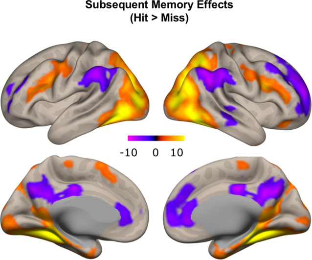
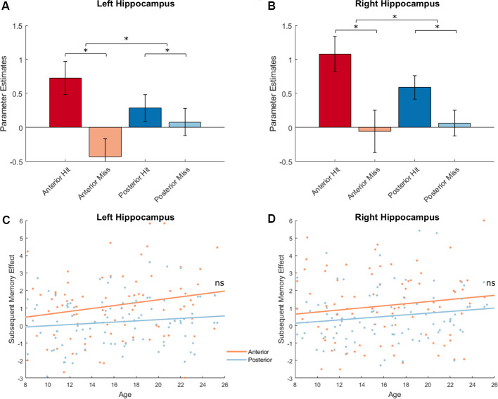
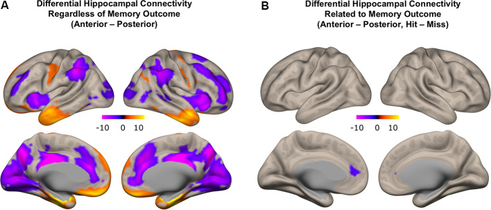
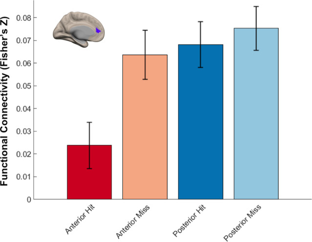
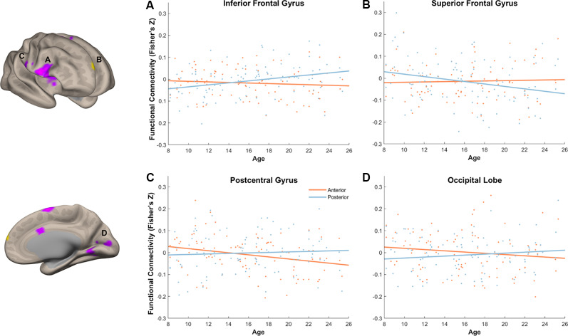
Neuroimaging evidence suggests that the development of the hippocampus, a brain structure critical for memory function, contributes to the improvements of episodic memory between middle childhood to adulthood. However, investigations on age differences in hippocampal activation and functional connectivity and their contributions to the development of memory have yielded mixed results. Given the known structural and functional heterogeneity along the long axis of the hippocampus, we investigated age differences in the activation and functional connectivity in hippocampal subregions with a cross-sectional sample of 96 participants ages 8-25 years. We found that anterior and posterior hippocampus supported memory formation, and there was overall stability in memory-related hippocampal activation with age. Without taking account of memory outcome, direct contrast between subregions showed higher functional connectivity of anterior, compared to the posterior hippocampus, with regions in the inferior frontal and lateral temporal lobes, and higher functional connectivity of posterior, compared to the anterior hippocampus, with regions in the medial and superior frontal, inferior parietal, and occipital lobes. A direct contrast between the memory-related connectivity patterns of anterior and posterior hippocampus identified a region in the medial frontal cortex, with which anterior and posterior hippocampus was differentially functionally connected. Finally, we identified age differences in memory-related differential hippocampal functional connectivity with several frontal and visual/sensory cortices, underscoring the importance of examining age differences in the patterns of hippocampal connectivity. Moreover, the specific patterns of differential anterior and posterior functional connectivity indicate an increase in the functional specialization along the long axis of the hippocampus and a dynamic shift in hippocampal connectivity patterns that supports memory development.
Keywords: MRI; anterior; connectivity; development; hippocampus; memory; posterior.
Copyright © 2020 Tang, Pruitt, Yu, Homayouni, Daugherty, Damoiseaux and Ofen.
Figures
Similar articles
-
Volume of the posterior hippocampus mediates age-related differences in spatial context memory and is correlated with increased activity in lateral frontal, parietal and occipital regions in healthy aging.Neuroimage. 2022 Jul 1;254:119164. doi: 10.1016/j.neuroimage.2022.119164. Epub 2022 Apr 4. Neuroimage. 2022. PMID: 35381338
-
Longitudinal Differences in Human Hippocampal Connectivity During Episodic Memory Processing.Cereb Cortex Commun. 2020;1(1):tgaa010. doi: 10.1093/texcom/tgaa010. Epub 2020 Apr 14. Cereb Cortex Commun. 2020. PMID: 32864613 Free PMC article.
-
Differential connectivity of perirhinal and parahippocampal cortices within human hippocampal subregions revealed by high-resolution functional imaging.J Neurosci. 2012 May 9;32(19):6550-60. doi: 10.1523/JNEUROSCI.3711-11.2012. J Neurosci. 2012. PMID: 22573677 Free PMC article.
-
Meta-analytic and functional connectivity evidence from functional magnetic resonance imaging for an anterior to posterior gradient of function along the hippocampal axis.Hippocampus. 2020 May;30(5):456-471. doi: 10.1002/hipo.23164. Epub 2019 Oct 7. Hippocampus. 2020. PMID: 31589003 Review.
-
Cortico-hippocampal systems involved in memory and cognition: the PMAT framework.Prog Brain Res. 2015;219:45-64. doi: 10.1016/bs.pbr.2015.04.001. Epub 2015 May 23. Prog Brain Res. 2015. PMID: 26072233 Review.
Cited by
-
The Analysis of Oxidative Stress Markers May Increase the Accuracy of the Differential Diagnosis of Alzheimer's Disease with and without Depression.Clin Interv Aging. 2021 Jun 16;16:1105-1117. doi: 10.2147/CIA.S310750. eCollection 2021. Clin Interv Aging. 2021. PMID: 34163154 Free PMC article.
-
Motifs of human high-frequency oscillations structure processing and memory of continuous audiovisual narratives.Sci Adv. 2025 Jul 25;11(30):eadv0986. doi: 10.1126/sciadv.adv0986. Epub 2025 Jul 25. Sci Adv. 2025. PMID: 40712018 Free PMC article.
-
Structural and functional sex differences in medial temporal lobe subregions at midlife.BMC Neurosci. 2024 Oct 25;25(1):55. doi: 10.1186/s12868-024-00905-9. BMC Neurosci. 2024. PMID: 39455948 Free PMC article.
-
Hippocampal anterior- posterior shift in childhood and adolescence.Prog Neurobiol. 2023 Jun;225:102447. doi: 10.1016/j.pneurobio.2023.102447. Epub 2023 Mar 24. Prog Neurobiol. 2023. PMID: 36967075 Free PMC article.
-
Exploring the late maturation of an intrinsic episodic memory network: A resting-state fMRI study.Dev Cogn Neurosci. 2024 Dec;70:101453. doi: 10.1016/j.dcn.2024.101453. Epub 2024 Sep 26. Dev Cogn Neurosci. 2024. PMID: 39368283 Free PMC article.
References
Grants and funding
LinkOut - more resources
Full Text Sources