. 2020 Jun 5:11:844.
doi: 10.3389/fphar.2020.00844. eCollection 2020.

Eye-Light on Age-Related Macular Degeneration: Targeting Nrf2-Pathway as a Novel Therapeutic Strategy for Retinal Pigment Epithelium

Affiliations

Eye-Light on Age-Related Macular Degeneration: Targeting Nrf2-Pathway as a Novel Therapeutic Strategy for Retinal Pigment Epithelium

Michele Catanzaro et al. Front Pharmacol. .

Abstract

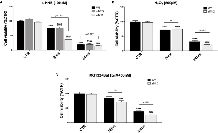
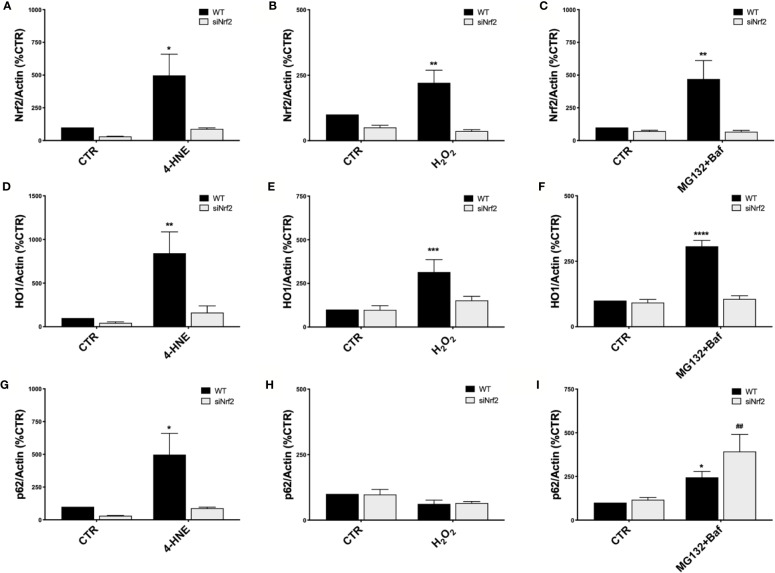
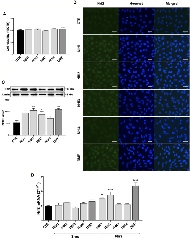
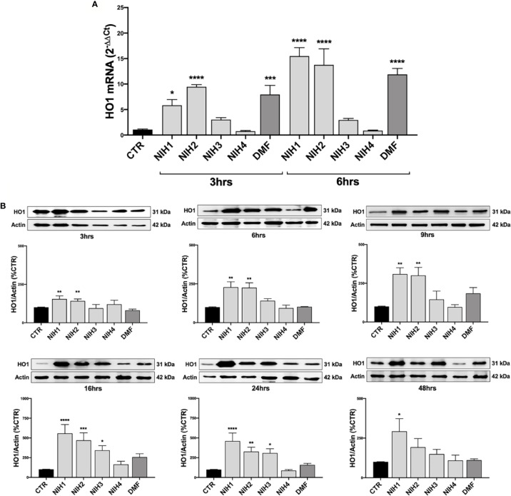
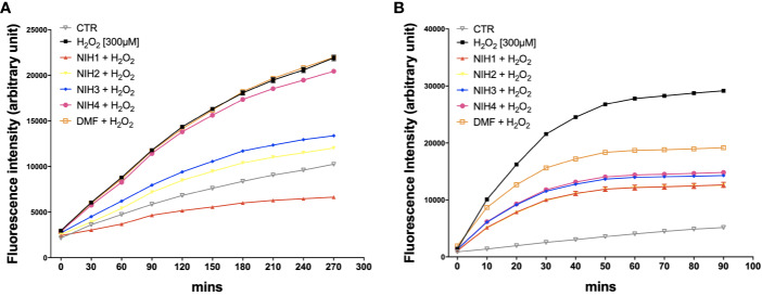
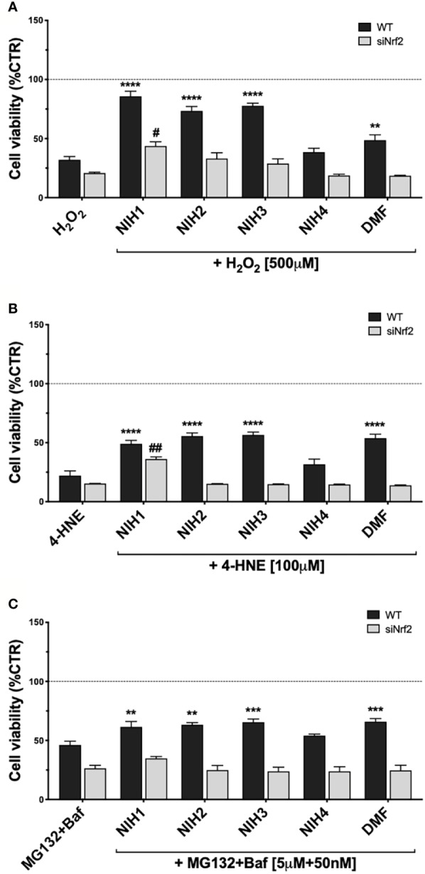
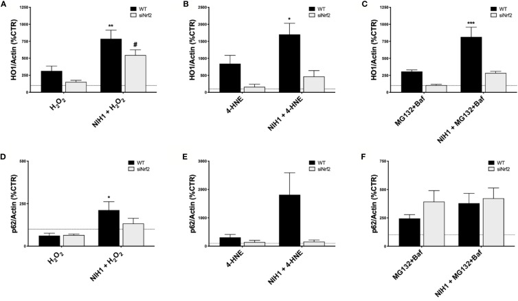
Age-related macular degeneration (AMD) is a common disease with a multifactorial aetiology, still lacking effective and curative therapies. Among the early events triggering AMD is the deterioration of the retinal pigment epithelium (RPE), whose fundamental functions assure good health of the retina. RPE is physiologically exposed to high levels of oxidative stress during its lifespan; thus, the integrity and well-functioning of its antioxidant systems are crucial to maintain RPE homeostasis. Among these defensive systems, the Nrf2-pathway plays a primary role. Literature evidence suggests that, in aged and especially in AMD RPE, there is an imbalance between the increased pro-oxidant stress, and the impaired endogenous detoxifying systems, finally reverberating on RPE functions and survival. In this in vitro study on wild type (WT) and Nrf2-silenced (siNrf2) ARPE-19 cells exposed to various AMD-related noxae (H2O2, 4-HNE, MG132 + Bafilomycin), we show that the Nrf2-pathway activation is a physiological protective stress response, leading downstream to an up-regulation of the Nrf2-targets HO1 and p62, and that a Nrf2 impairment predisposes the cells to a higher vulnerability to stress. In search of new pharmacologically active compounds potentially useful for AMD, four nature-inspired hybrids (NIH) were individually characterized as Nrf2 activators, and their pharmacological activity was investigated in ARPE-19 cells. The Nrf2 activator dimethyl-fumarate (DMF; 10 μM) was used as a positive control. Three out of the four tested NIH (5 μM) display both direct and indirect antioxidant properties, in addition to cytoprotective effects in ARPE-19 cells under pro-oxidant stimuli. The observed pro-survival effects require the presence of Nrf2, with the exception of the lead compound NIH1, able to exert a still significant, albeit lower, protection even in siNrf2 cells, supporting the concept of the existence of both Nrf2-dependent and independent pathways mediating pro-survival effects. In conclusion, by using some pharmacological tools as well as a reference compound, we dissected the role of the Nrf2-pathway in ARPE-19 stress response, suggesting that the Nrf2 induction represents an efficient defensive strategy to prevent the stress-induced damage.

Keywords: HO1; Nrf2; age-related macular degeneration (AMD); cytoprotection; oxidative stress; p62; pharmacological modulation; retinal pigment epithelium (RPE).


Figures

Figure 1
Figure 1
The Nrf2-impaired ARPE-19 cells are more susceptible to stress than their WT counterpart. Cell viability was assessed by a fluorometric assay (PrestoBlue®) and results are expressed as mean of percentages ± SEM. (A) WT, siNEG, and siNrf2 ARPE-19 cells were treated with either solvent (DMSO, CTR) or 4-HNE [100 μM] for 8 and 24 h. Dunnett’s multiple comparisons test; ****p < 0.0001 versus WT CTR; §§§§p < 0.0001 versus siNEG CTR; and ####p < 0.0001 versus siNrf2 CTR; ns = non significant; n = 5–8. (B) WT and siNrf2 ARPE-19 cells were treated with either solvent (DMSO, CTR) or H2O2 [500 μM] for 8 and 24 h. Sidak’s multiple comparisons test; ****p = 0.0001 versus WT CTR; ####p < 0.0001 versus siNrf2 CTR; n = 6–8. (C) WT and siNrf2 ARPE-19 cells were treated with either solvent (DMSO, CTR) or MG132 + Bafilomycin [5 μM + 50 nM] for 24 and 48 h. Sidak’s multiple comparisons test; ****p < 0.0001 versus WT CTR; ###p < 0.001 and ####p < 0.0001 versus siNrf2 CTR; ns = non significant; n = 5–9.
Figure 2
Figure 2
Nrf2-pathway activation in wild type ARPE-19 cells under different stress conditions. Both WT and siNrf2 ARPE-19 cell lines were treated for 6 h with either solvent (DMSO, CTR), 4-HNE [50 μM] (A, D, G), H2O2 [300 μM] (B, E, H), or MG132 + Bafilomycin [5 μM + 50 nM] (C, F, I). Total cellular homogenates were examined by Western blot using an antibody against Nrf2 (AC), HO1 (DF), and p62 (GI); Actin content was used as a loading control to normalize the data. Results are expressed as percentage means ± SEM. Sidak’s multiple comparisons test; *p < 0.05, **p < 0.01, ***p < 0.001, and ****p < 0.0001 versus WT CTR; ##p < 0.01 versus siNrf2 CTR; n = 4–6.
Figure 3
Figure 3
Pharmacological activation of Nrf2 protein by NIH in ARPE-19 cells. (A) Tolerability of NIH in ARPE-19 cells. ARPE-19 cells were treated for 48 h with solvent (DMSO, CTR), NIH1, NIH2, NIH3 and NIH4 [5 μM], or DMF [10 μM], and their viability was assessed by MTT assay. Results are expressed as mean of percentages ± SEM (CTR: 100%); Dunnett’s multiple comparisons test; no statistical differences versus CTR; n = 3–5. (BC) Evaluation of NIH-induced Nrf2 nuclear translocation. ARPE-19 cells were treated for 3 h with either solvent (DMSO, CTR), NIH1, NIH2, NIH3, NIH4 [5 μM], or DMF [10 μM].(B) Immunocytochemistry for Nrf2 protein localization was performed by using a green fluorescent-conjugated secondary antibody. Nuclei (blue) were stained by Hoechst. Scale bar: 20 μm. (C) Nuclear fraction was isolated and examined by Western blot using an antibody against Nrf2. Lamin A content was used as a loading control to normalize the data. Results are expressed as means of Nrf2/Lamin A (ratio × 1000) ± SEM. Dunnett’s multiple comparisons test; *p<0.05 and **p<0.01 versus CTR; n=5. (D) Study of Nrf2 mRNA expression following NIH treatment. Total Nrf2 mRNA levels were measured by RT-qPCR in ARPE-19 cells exposed to either solvent (DMSO, CTR), NIH1, NIH2, NIH3, NIH4 [5 μM], or DMF [10 μM] for 3 and 6 h. GAPDH mRNA content was used as a housekeeping to normalize the data. Results are expressed as means of 2−ΔΔCt ± SEM. Dunnett’s multiple comparisons test; **p <0.01 and ****p <0.0001 versus CTR; n = 3.
Figure 4
Figure 4
The NIH-mediated increase of HO1 expression vary among molecules. (A) Total HO1 mRNA levels were measured by RT-qPCR in ARPE-19 cells exposed to either solvent (DMSO, CTR), NIH1, NIH2, NIH3 and NIH4 [5 μM], or DMF [10 μM], for 3 and 6 h. GAPDH mRNA content was used as a housekeeping to normalize the data. Results are expressed as means of 2−ΔΔCt ± SEM. Dunnett’s multiple comparisons test; *p <0.05, ***p <0.001 and ****p <0.0001 versus CTR; n = 3. (B) ARPE-19 cells were exposed to either solvent (DMSO, CTR), NIH1, NIH2, NIH3, NIH4 [5 μM], or DMF [10 μM], for increasing times (up to 48 h), and total homogenates were examined by Western blot using an antibody against HO1. Actin was used as a loading control to normalize the data. Results are expressed as mean percentages ± SEM. Dunnett’s multiple comparisons test; *p <0.05, **p <0.01; ***p <0.001 and ****p <0.0001 versus CTR; n = 5.
Figure 5
Figure 5
NIH are endowed of anti-oxidant properties. (A) ARPE-19 cells were exposed to H2O2 [300 μM] ± NIH1, NIH2, NIH3 and NIH4 [5 μM], or DMF [10 μM] for 4.5 h. Control cells were exposed only to solvent (DMSO). ROS levels were measured every 30 min by the fluorometric 2′,7′-Dichlorofluorescin diacetate (DCF-DA) assay. Results are expressed as mean ± SEM. Fluorescence intensity for the NIH1, NIH2, NIH3, is significantly different from H2O2 at any time starting from 30 min with p <0.001. Dunnett’s multiple comparisons test versus H2O2; n = 4. (B) ARPE-19 cells were pre-treated for 24 h with either solvent (DMSO, CTR), NIH1, NIH2, NIH3, NIH4 [5 μM], or DMF [10 μM], and then exposed to H2O2 [300 μM] for 1.5 h. ROS levels were measured every 10 min by the DCF-DA assay. Results are expressed as mean ± SEM. Fluorescence intensity for all the NIH and DMF is significant at any time starting from 10 min with p <0.0001. Dunnett’s multiple comparisons test versus H2O2; n = 5.
Figure 6
Figure 6
Study of NIH-mediated protection in WT and siNrf2 ARPE-19 cells under different stress. Cell viability was assessed by a fluorometric assay (PrestoBlue ®) in WT and siNrf2 ARPE-19 cells pre-treated for 24 h with either solvent (DMSO, CTR), NIH1, NIH2, NIH3, NIH4 [5 μM], or DMF [10 μM], and then exposed to either H2O2 [500 μM, for 24 h] (A), 4-HNE [100 μM, for 24 h] (B), or MG132 + Bafilomycin [5 μM + 50 nM, for 48 h] (C). Results are expressed as mean of percentages ± SEM in comparison with control (dot line, 100%). Dunnett’s multiple comparisons test; *p <0.05, **p < 0.01, ***p < 0.001, and ****p < 0.0001 versus WT stress; #p < 0.05 and ##p < 0.01 versus siNrf2 stress; n = 5–10.
Figure 7
Figure 7
NIH1-mediated modulation of HO1 and p62 protein expression in stressed WT and siNrf2 ARPE-19 cells. Both WT and siNrf2 ARPE-19 cell lines were pre-treated for 24 h with either solvent (DMSO, CTR), or NIH1 [5 μM], and then stressed, or not, for 6 h with H2O2 [300 μM] (A, D), 4-HNE [50 μM] (B, E), or MG132 + Bafilomycin [5 μM + 50 nM] (C, F). Total cellular homogenates were examined by Western blot using antibodies against HO1 (AC) and p62 (DF); Actin content was used as a loading control to normalize the data. Results are expressed as percentage means ± SEM in comparison with control (dot line, 100%). Sidak’s multiple comparisons test; *p < 0.05, **p < 0.01; and ***p < 0.001 versus WT stress; #p < 0.05 versus siNrf2 stress; n = 4–6.

References

    1. Abokyi S., To C.-H., Lam T. T., Tse D. Y. (2020). Central Role of Oxidative Stress in Age-Related Macular Degeneration: Evidence from a Review of the Molecular Mechanisms and Animal Models. Oxid. Med. Cell. Longev. 2020, 1–19. 10.1155/2020/7901270 - DOI - PMC - PubMed
    1. Amadio M., Scapagnini G., Laforenza U., Intrieri M., Romeo L., Govoni S., et al. (2008). Post-transcriptional regulation of HSP70 expression following oxidative stress in SH-SY5Y cells: the potential involvement of the RNA-binding protein HuR. Curr. Pharm. Des. 14, 2651–2658. 10.2174/138161208786264052 - DOI - PubMed
    1. Amadio M., Scapagnini G., Davinelli S., Calabrese V., Govoni S., Pascale A. (2014). Involvement of ELAV RNA-binding proteins in the post-transcriptional regulation of HO-1. Front. Cell. Neurosci. 8, 459. 10.3389/fncel.2014.00459 - DOI - PMC - PubMed
    1. Amadio M., Govoni S., Pascale A. (2016). Targeting VEGF in eye neovascularization: What’s new? Pharmacol. Res. 103, 253–269. 10.1016/j.phrs.2015.11.027 - DOI - PubMed
    1. Ambati J., Fowler B. J. (2012). Mechanisms of Age-Related Macular Degeneration. Neuron 75, 26–39. 10.1016/j.neuron.2012.06.018 - DOI - PMC - PubMed

LinkOut - more resources