Review
. 2020 Jun 5:11:853.
doi: 10.3389/fphar.2020.00853. eCollection 2020.

Transgenic Rabbit Models in Proarrhythmia Research

Affiliations
Review

Transgenic Rabbit Models in Proarrhythmia Research

István Baczkó et al. Front Pharmacol. .

Abstract

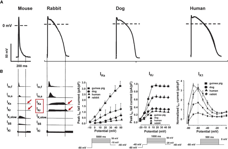
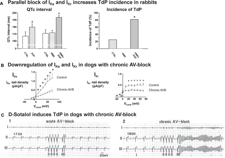
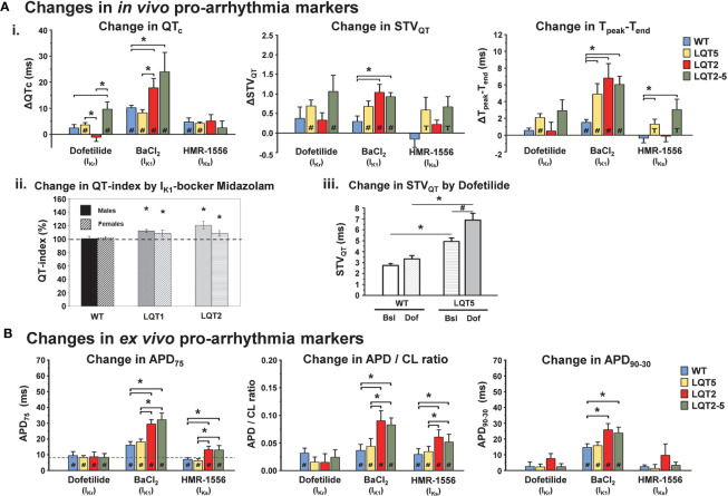
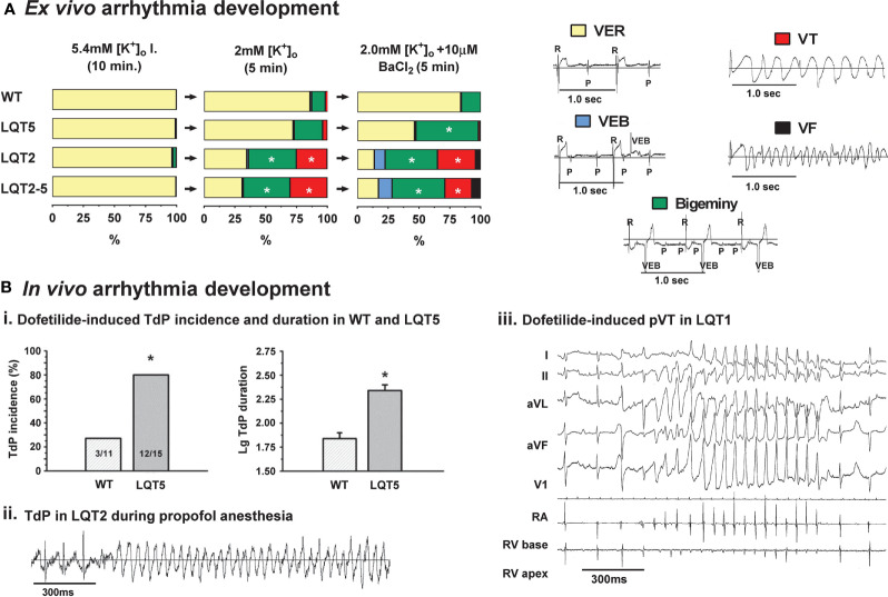
Drug-induced proarrhythmia constitutes a potentially lethal side effect of various drugs. Most often, this proarrhythmia is mechanistically linked to the drug's potential to interact with repolarizing cardiac ion channels causing a prolongation of the QT interval in the ECG. Despite sophisticated screening approaches during drug development, reliable prediction of proarrhythmia remains very challenging. Although drug-induced long-QT-related proarrhythmia is often favored by conditions or diseases that impair the individual's repolarization reserve, most cellular, tissue, and whole animal model systems used for drug safety screening are based on normal, healthy models. In recent years, several transgenic rabbit models for different types of long QT syndromes (LQTS) with differences in the extent of impairment in repolarization reserve have been generated. These might be useful for screening/prediction of a drug's potential for long-QT-related proarrhythmia, particularly as different repolarizing cardiac ion channels are impaired in the different models. In this review, we summarize the electrophysiological characteristics of the available transgenic LQTS rabbit models, and the pharmacological proof-of-principle studies that have been performed with these models-highlighting the advantages and disadvantages of LQTS models for proarrhythmia research. In the end, we give an outlook on potential future directions and novel models.

Keywords: K+-channel blocker; cardiac repolarization reserve; drug-induced proarrhythmia; long QT syndrome; proarrhythmia safety screening; transgenic LQTS rabbit.


Figures

Figure 1
Figure 1
Species differences in repolarizing ion currents. (A) Representative action potential (AP) recordings [modified from (Rudy et al., 2008; Blenck et al., 2016)] and (B) schematic illustration of the main repolarizing ion currents in mouse, rabbit, dog, and human cardiomyocytes [modified from (Nerbonne et al., 2001)]. Panel (C) shows species differences in IKs, IKr, and IK1 in rabbit, dog, and human.
Figure 2
Figure 2
Pro-arrhythmia detection with acquired LQT rabbit and chronic AV-block dog models. (A) Left panel: The QTc interval was significantly prolonged by the IKr blocker dofetilide and was further prolonged by the combination of dofetilide and the IKr blocker HMR1556 given in any order, while IKs block alone did not prolong the QTc in anesthetized rabbits. Right panel: IKr block caused TdP in some of the animals, IKs block alone did not induce TdP in anesthetized rabbits. Importantly, combined acute pharmacological block of IKs and IKr, given in any order, caused a significant increase in TdP incidence. #p < 0.05 vs. baseline control in the same group; *p < 0.05 vs. dofetilide. N=7-11 animals/group. CTRL: Control, DOF: Dofetilide, HMR: HMR-1556. Modified from (Lengyel et al., 2007a). (B) In right ventricular cardiomyocytes isolated from dogs with chronic AV-block and marked myocardial hypertrophy, IKs tail current density was reduced by approximately 55% (n=9), while IKr tail current density was reduced by 45% (n=14) compared to those isolated from control animals. *p < 0.05 vs. control myocytes. Modified from (Volders et al., 1999). (C) Standard bipolar ECG lead recordings illustrating a marked difference in the arrhythmogenic response to administration of the selective IKr blocker D-sotalol in dogs with acute (left panel) and chronic AV-block (right panel). The pacing protocol involved a basic pacing cycle length (CL) of 1730 ms in acute AV block, and 1800 ms spontaneous idioventricular CL in chronic AV-block. Arrhythmia provocation included a short/long/short sequence at 600 ms CL for 4 beats followed by 2 paced beats with 1200 and 350 ms cycle lengths. No TdP was observed in acute AV-block dogs, while 60% of animals with chronic AV-block exhibited TdP. Modified from (Vos et al., 1998).
Figure 3
Figure 3
Baseline electrical characteristics of transgenic LQTS rabbits. (A) Upper panel: IV-curves of IKs steady (left)/tail (right) and IKr steady (left)/tail (right) in cardiomyocytes isolated from wild-type (WT), LQT1, and LQT2 rabbit hearts, indicating loss of IKs in LQT1 and loss of IKr in LQT2 [modified from (Brunner et al., 2008)]. Lower panel: IV-curves of IKs in absence and presence of 5 µM forskolin in WT or transgenic LQT5 rabbit ventricular myocytes. Bar diagrams illustrate a reduced deactivation time constant in transgenic LQT5 ventricular myocytes [modified from (Major et al., 2016)]. (B) Representative ECG tracings indicating differences in QT interval in WT, LQT1, LQT2, and LQT5 rabbits [ECG from WT, LQT1 and LQT2 modified from (Brunner et al., 2008)]. (C) QT/RR relationship assessed with telemetric ECG in free moving rabbits: WT, LQT1, and LQT2 in upper panel [modified from (Brunner et al., 2008)], in lower panel in WT and LQT5 [modified from (Hornyik et al., 2020)]. (D) ECG and blood pressure tracing of LQT2 rabbit with spontaneous ventricular torsade-de-pointes (TdP) tachycardia [modified from (Brunner et al., 2008)]. *p < 0.05 vs. WT.
Figure 4
Figure 4
Mechanisms of arrhythmogenesis in transgenic LQT2 rabbits. (A) Upper panel (i): Bar graphs of action potential durations (APD) and dispersion of APD (ΔAPD) in Langendorff-perfused hearts of WT, LQT1, and LQT2 rabbits indicate longer APD and increased APD dispersion in LQT2 hearts, *p < 0.05 [modified from (Brunner et al., 2008)]. Lower panel (ii): optical mapping APD map (left; indicated are isochrones of APD, in ms) and activation maps (right) of initial beats of ventricular tachycardia/fibrillation (VF) in an estradiol-treated LQT2 heart indicate reentry formation (indicated by red arrows) in line with the regional APD heterogeneities [modified from (Odening et al., 2012b)]. (B) Representative trace of Ca2+ oscillations and early afterdepolariaztion (EAD) in estradiol-treated LQT2 rabbit heart after AV-node ablation and bolus application of isoproterenol (140 nM). Black line indicates changes in voltage fluorescence signal (Vm); red line indicates changes in Ca2+ signal [modified from (Odening et al., 2012b)].
Figure 5
Figure 5
Pro-arrhythmic drug-effects in transgenic LQTS rabbit models: changes in pro-arrhythmia markers. (A) Changes in in vivo pro-arrhythmia markers. (i.) Bar graphs show changes in QTc, STVQT, and Tpeak-Tend in anaesthetized animals after i.m. injection of IKr, IK1, and IKs-blockers dofetilide, BaCl2, HMR-1556, respectively in WT, LQT2, LQT5, and LQT2–5 rabbits [modified from (Hornyik et al., 2020)]. *p < 0.05 inter-genotype comparison, #p < 0.05 vs. baseline, T trend p < 0.1 vs. baseline. (ii.) IK1-blocker midazolam-induced change in heart-rate corrected QT-index in free-moving male (solid bars) and female (hatched bars) WT, LQT1, and LQT2 rabbits is also shown. The dashed line represents the mean QT indexes in free-moving rabbits obtained with the genotype-specific correction formula (=100%) [modified from (Odening et al., 2008)]. *p < 0.05 vs. free-moving rabbits of the same genotype. (iii.) Bar graphs indicate IKr-blocker dofetilide-induced increase in STVQT in WT and LQT5 animals [modified from (Major et al., 2016)]. Bsl, baseline; Dof, dofetilide.*p < 0.05 inter-genotype comparison, #p < 0.05 vs. baseline. (B) Changes in ex vivo pro-arrhythmia markers. Bar graphs of changes in action potential duration (ΔAPD75), action potential duration/stimulation cycle length ratio (ΔAPD/CL ratio and action potential triangulation (ΔAPD90-30) after 10 min perfusion with IKr, IK1 and IKs-blockers dofetilide, BaCl2 and HMR-1556, respectively in WT, LQT2, LQT5, and LQT2–5 rabbits. *p < 0.05 inter-genotype comparison, #p < 0.05 vs. baseline [modified from (Hornyik et al., 2020)].
Figure 6
Figure 6
Pro-arrhythmic drug-effects in transgenic LQTS rabbit models: arrhythmia development. (A) Ex vivo arrhythmia development. Graphs indicating the duration (% of perfusion time) of arrhythmias provoked by perfusing the hearts with low [K+]o (2 mM) KH, or with combined low [K+]o (2 mM) KH and IK1-blocker BaCl2 (10 µM) in WT, LQT5, LQT2 and LQT2–5 animals. Inlets show representative ECG recordings of ventricular escape rhythm (VER), ventricular extra beats (VEB), bigeminy, ventricular tachycardia (VT) and ventricular fibrillation (VF) [modified from (Hornyik et al., 2020)]. *p < 0.05 inter-genotype comparison. (B) In vivo arrhythmia development. (i.) Bar graphs of dofetilide-induced incidence (in %) and log duration of torsade de pointes (TdP) tachycardia in WT and LQT5 rabbits [modified from (Major et al., 2016)]. *p < 0.05 inter-genotype comparison. (ii.) Episode of polymorphic TdP tachycardia in female LQT2 rabbit during propofol anesthesia (acquired with telemetric ECG) [modified from (Odening et al., 2008)]. (iii.) Episode of dofetilide-induced pVT in male LQT1 rabbit during episode of alternating AV 2:1/3:1 block [modified from (Odening et al., 2010)].

Similar articles

Cited by

References

    1. Amos G. J., Abrahamsson C., Duker G., Hondeghem L., Palmer M., Carlsson L. (2001). Potassium and calcium current blocking properties of the novel antiarrhythmic agent H 345/52: implications for proarrhythmic potential. Cardiovasc. Res. 49, 351–360. 10.1016/S0008-6363(00)00259-5 - DOI - PubMed
    1. Anderson M. E. (2006). QT interval prolongation and arrhythmia: an unbreakable connection? J. Intern. Med. 259, 81–90. 10.1111/j.1365-2796.2005.01580.x - DOI - PubMed
    1. Ando F., Kuruma A., Kawano S. (2011). Synergic effects of beta-estradiol and erythromycin on hERG currents. J. Membr. Biol. 241, 31–38. 10.1007/s00232-011-9360-z - DOI - PubMed
    1. Antoons G., Oros A., Beekman J. D., Engelen M. A., Houtman M. J., Belardinelli L., et al. (2010). Late na(+) current inhibition by ranolazine reduces torsades de pointes in the chronic atrioventricular block dog model. J. Am. Coll. Cardiol. 55, 801–809. 10.1016/j.jacc.2009.10.033 - DOI - PubMed
    1. Antzelevitch C. (2007). Role of spatial dispersion of repolarization in inherited and acquired sudden cardiac death syndromes. Am. J. Physiol. Heart Circ. Physiol. 293, H2024–H2038. 10.1152/ajpheart.00355.2007 - DOI - PMC - PubMed