. 2020 Jun 3:11:438.
doi: 10.3389/fphys.2020.00438. eCollection 2020.

Mechanisms Associated to Nitroxyl (HNO)-Induced Relaxation in the Intestinal Smooth Muscle

Affiliations

Mechanisms Associated to Nitroxyl (HNO)-Induced Relaxation in the Intestinal Smooth Muscle

Mirko Gastreich-Seelig et al. Front Physiol. .

Abstract

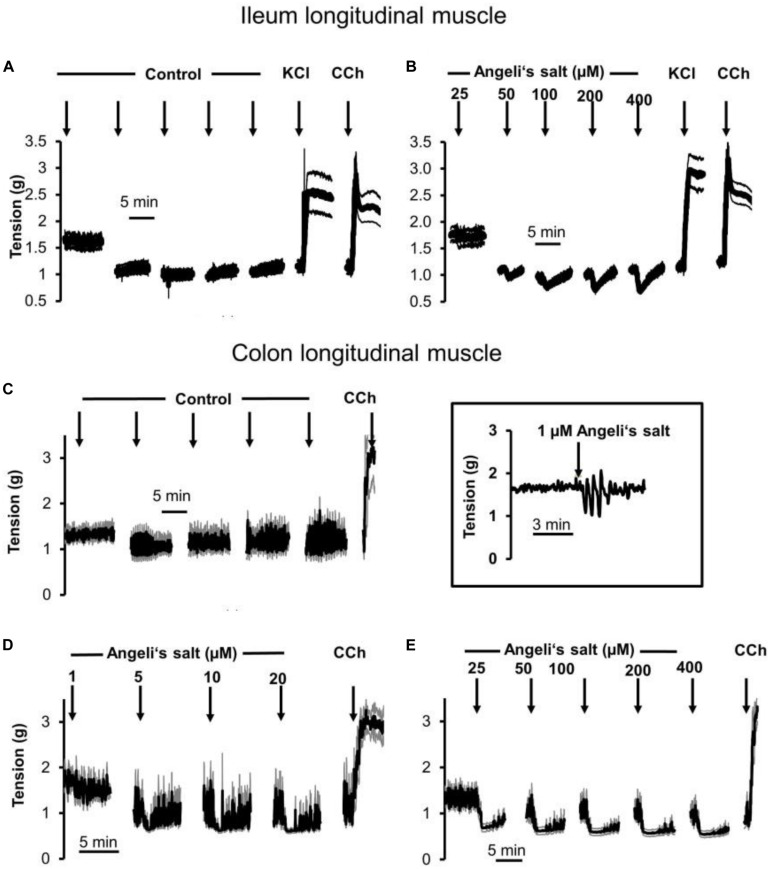
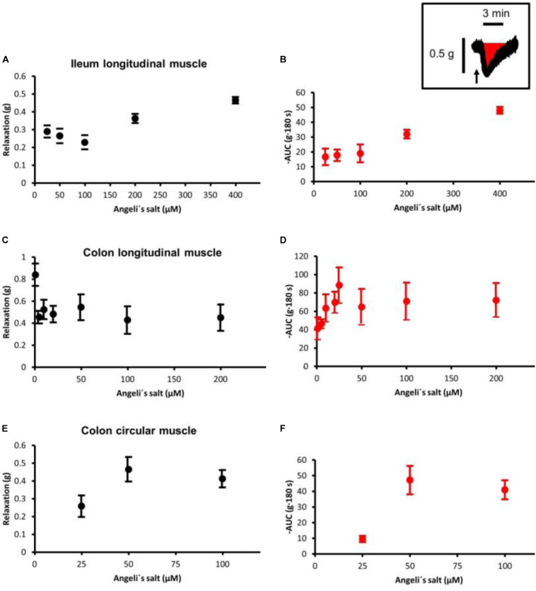
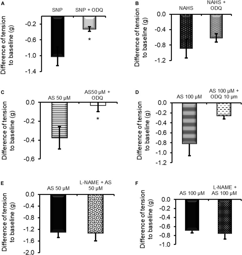
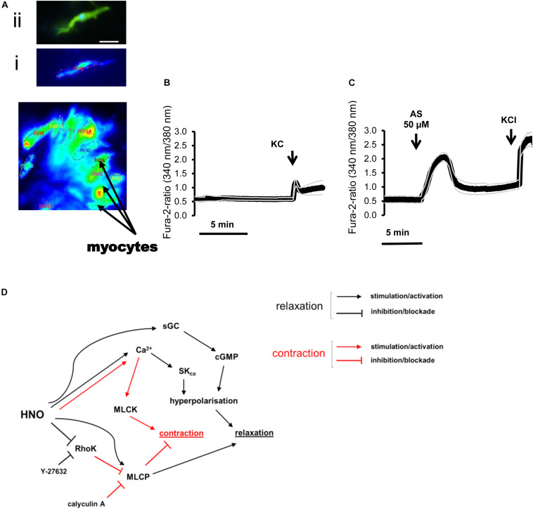
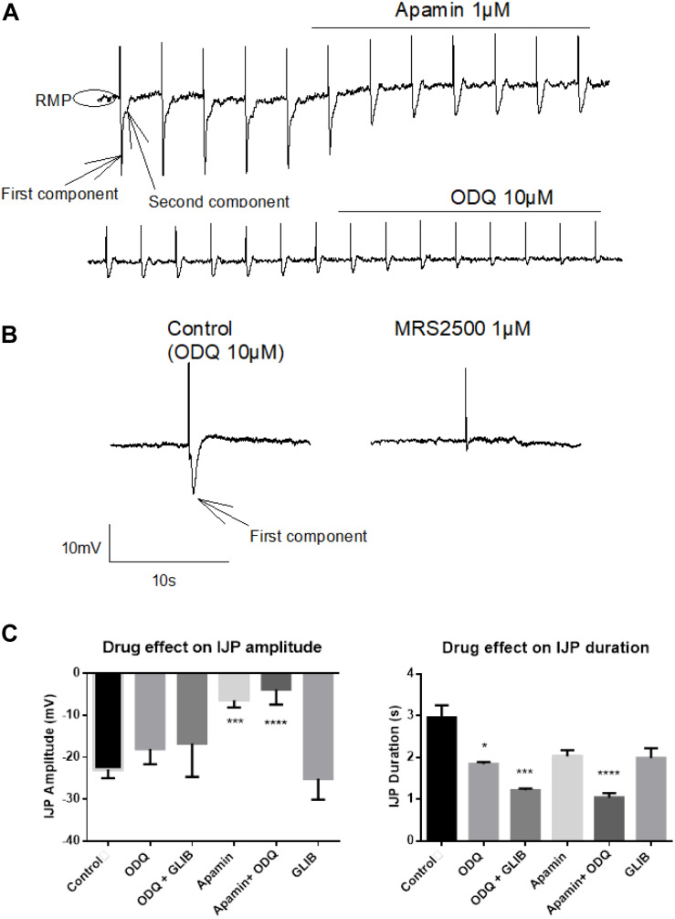
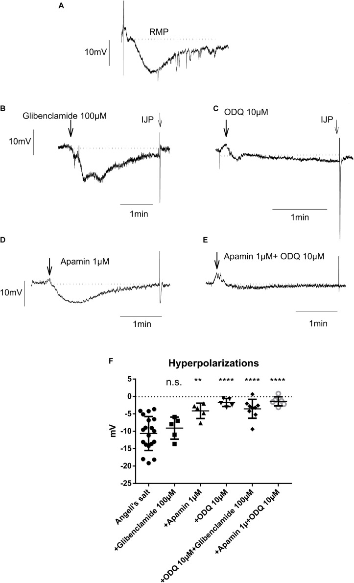
The pharmacological properties of nitroxyl (HNO) donors in the gastrointestinal tract are unknown. We investigated the properties of this molecule in the regulation of gastrointestinal contractility focusing on its possible interaction with other gaseous signaling molecules such as NO and H2S. Organ bath, Ca2+ imaging, and microelectrode recordings were performed on rat intestinal samples, using Angeli's salt as HNO donor. Angeli's salt caused a concentration-dependent relaxation of longitudinal or circular muscle strips of the ileum and the proximal colon. This relaxation was strongly inhibited by the Rho-kinase inhibitor Y-27632 (10 μM), by the reducing agent DTT or by the inhibitor of soluble guanylate cyclase (sGC) ODQ (10 μM) alone or in combination with the inhibitors of the endogenous synthesis of H2S β-cyano-L-alanine (5 mM) and amino-oxyacetate (5 mM). Preventing endogenous synthesis of NO by the NO synthase inhibitor L-NAME (200 μM) did not affect the relaxation induced by HNO. HNO induced an increase in cytosolic Ca2+ concentration in colonic myocytes. It also elicited myocyte membrane hyperpolarization that amounted to -10.6 ± 1.1 mV. ODQ (10 μM) and Apamin (1 μM), a selective inhibitor of small conductance Ca2+-activated K+ channels (SKca), strongly antagonized this effect. We conclude that HNO relaxes the gastrointestinal tract musculature by hyperpolarizing myocytes via activation of the sGC/cGMP pathway similarly to NO, not only inhibiting the RhoK and activating MLCP as do both NO and H2S but also increasing cytosolic Ca2+ for activation of SK C a contributing to hyperpolarization.

Keywords: Ca2+; gasotransmitter; membrane potential; motility; nitroxyl (HNO); small conductance Ca2+-activated K+ channels (SKca); soluble guanylate cyclase.


Figures

FIGURE 1
FIGURE 1
Relaxation induced by different concentrations of Angeli’s salt (1–400 μM, arrows) on longitudinal segments of (B) rat ileum or (D,E) proximal colon and (A) or (C) time-dependent control experiments, where only the solvent of Angeli’s salt was administered. The inset (middle) shows an original tracing of an individual colonic longitudinal muscle strip with a transient relaxation induced by 1 μM Angeli’s salt, which is hard to recognize in the ensemble average depicted in (D) resulting from the averaging of eight muscle strips responding asynchronously with a short relaxation induced by the HNO donor. Line interruptions are caused by omitting washing periods of about 10 min, where the content of the organ bath was exchanged three times before the next concentration of Angeli’s salt was administered. KCl (30 mM) and or carbachol (CCh; 10 μM) were used to check tissue viability. Values are means of individual tracings (black lines) ± SEM (gray lines), n = 5–11. For statistics (B,D,E) are plotted in Figures 3A–D, respectively.
FIGURE 2
FIGURE 2
The HNO donor Angeli’s salt (25–100 μM, arrows) induces a relaxation of circular segments of (B) rat proximal colon compared to a (A) time-dependent control, where only the solvent of Angeli’s salt was administered. Line interruptions are caused by omitting washing periods of about 10 min, where the content of the organ bath was exchanged three times before the next concentration of Angeli’s salt was administered. KCl (30 mM) and carbachol (CCh; 10 μM) were used to check tissue viability. Data in (A,B) are means of individual tracings (black lines) ± SEM (gray lines), n = 5–6. (C,D) are higher magnifications of 5-min intervals of an individual muscle preparation contained in the respective ensemble averages in (A,B). For statistics (B), is plotted in Figures 3E,F.
FIGURE 3
FIGURE 3
Concentration-dependent relaxation induced by Angeli’s salt in (A,B) ileal longitudinal muscle, (C,D) colonic longitudinal, and (E,F) colonic circular muscle. The relaxing effect is either expressed as maximal reduction in muscle tone (Δg; difference to baseline just prior to administration of the drug (A,C,E) or reduction in the area under the curve (AUC) over a 3-min period compared to the 3-min period just prior to administration of Angeli’s salt, as illustrated by the schematic inset where the arrow marks the administration of Angeli’s salt. Concentration–response curves in the colonic longitudinal muscle was constructed from two independent series of experiments (see Figure 1), in which the effect of 1–20 μM and 25–400 μM Angeli’s salt was tested, respectively. Values are means ± SEM, n = 6–11.
FIGURE 4
FIGURE 4
Inhibition of Rho kinases by Y-27632 (10 μM) prevents the contraction induced by CCh (10 μM) of longitudinal muscle strips from (A) proximal colon and reduces the relaxation induced by HNO donor Angeli’s salt (AS). Data in (B) are expressed as difference between the muscle tone just prior to administration of Angeli’s salt and the muscle tone averaged over a 1-min period starting 1 min after administration of the HNO. Potentiating (C) the contractile action of the cholinergic system with calyculin A (100 nM) strengthens or weakens the (D) relaxing action of HNO. Values are means ± SEM, n = 7–12. *P < 0.05, ***P < 0.001 vs. control in the absence of the corresponding blocker (Mann–Whitney U-test). For statistics, see text.
FIGURE 5
FIGURE 5
(A,C) The relaxing effect of the nitric oxide donor sodium nitroprusside (SNP; 1 mM) and 50 μM Angeli’s salt (AS) was significantly inhibited in the presence of the sGC inhibitor ODQ (10 μM). (D) When the concentration of Angeli’s salt was increased to 100 μM, this inhibition failed to reach significance. (B) Relaxation induced by the H2S donor sodium hydrogen sulfide (NaHS; 100 μM) was unaffected by ODQ. (E,F) Inhibition of NOS with L-NAME (200 μM) did not affect HNO-induced relaxation. n = 5–9. *P < 0.05, vs. control in the absence ODQ (Mann–Whitney U-test). For statistics, see text.
FIGURE 6
FIGURE 6
(A–C) Changes in the fura-2 ratio evoked by Angeli’s salt (50 μM) in isolated rat colonic myocytes and (D) mechanisms underlying relaxation. (A) Photograph of isolated myocytes loaded with fura-2. Insets show (i) a fura-2 loaded myocyte that responded to Angeli’s salt and the same cell (ii) after staining of actin filament with phalloidin (green) and nucleus with 4′,6-diamidino-2-phenylindole (DAPI) (blue). Scale bar, 50 μm. (C) Angeli’s salt induces an increase in the fura-2 ratio compared to (B) a time-dependent control. KCl (30 mM) was used for cell viability control. Values are given as means (symbols) ± SEM (parallel continuous lines), n = 6 for the time-dependent control and n = 13 for the test group with the HNO donor. For statistics, see text. (D) HNO may induce relaxation either directly by activating MLCP or indirectly inhibiting the RhoK. Additional increase in cytosolic Ca2+ concentration activates SKca for hyperpolarization and relaxation. Blocking RhoK with Y-27632 shunts HNO effects, inducing a shift toward Ca2+-mediated responses, which also activates MLCK for contraction. The main mechanism for HNO-induced relaxation consists in hyperpolarizing myocytes via activation of sGC/cGMP.
FIGURE 7
FIGURE 7
Identification of the fast inhibitory junction potential (IJPf) and sustained IJP (IJPs) evoked by electrical field stimulation (EFS) under non-adrenergic non-cholinergic (NANC) conditions. (A) Apamin blocks the IJPf [first component in (A)], while ODQ blocks the IJPs [second component in (A)]. Apamin induces a shift of the resting membrane potential (RMP) to more positive values. (A) The IJPf isolated after suppression of the IJPs by ODQ was also sensitive against MRS2500 (B). (C) The amplitude of the IJP was strongly inhibited by apamin; this inhibition was not enhanced, when, in addition, ODQ was administered. In contrast, the duration of the IJP was significantly reduced by ODQ; this effect was significantly enhanced when, in addition, glibenclamide or apamin was present. Concentrations of drugs were apamin 1 μM, glibenclamide 100 μM, and ODQ 10 μM. Values in (C) are means ± SEM, n = 3–12. *P < 0.05, ***P < 0.001, ****P < 0.0001, vs. control in the absence of any drug (analysis of variance followed by Bonferroni test). For statistics, see text.
FIGURE 8
FIGURE 8
The hyperpolarization caused by the HNO donor Angeli’s salt (25 μM; A, n = 19) is insensitive to glibenclamide (B,F, n = 5), but sensitive to apamin (D,F, n = 5) or highly sensitive to ODQ alone (C,F, n = 5) or in combination with apamin (E,F, n = 6) or glibenclamide (F, n = 9). The discontinuous horizontal lines represent the resting membrane potential (RMP). Values are means ± SD, n values refer to (F), while (A–E) are representative original tracings. **P < 0.01, ****P < 0.0001, vs. control in the absence of any drug (analysis of variance followed by Bonferroni test). For statistics, see text.

Similar articles

Cited by

References

    1. An W., Ryan L. S., Reeves A. G., Bruemmer K. J., Mouhaffel L., Gerberich J. L., et al. (2019). A Chemiluminescent Probe for HNO quantification and real-time monitoring in living cells. Angew. Chem. Int. Ed. 58 1361–1365. 10.1002/anie.201811257 - DOI - PMC - PubMed
    1. Baker S. A., Hennig G. W., Ward S. M., Sanders K. M. (2015). Temporal sequence of activation of cells involved in purinergic neurotransmission in the colon. J. Physiol. 593 1945–1963. 10.1113/jphysiol.2014.287599 - DOI - PMC - PubMed
    1. Bartberger M. D., Liu W., Ford E., Miranda K. M., Switzer C., Fukuto J. M., et al. (2002). The reduction potential of nitric oxide (NO) and its importance to NO biochemistry. Proc. Natl. Acad. Sci. U.S.A. 99:10958. 10.1073/pnas.162095599 - DOI - PMC - PubMed
    1. Boeckxstaens G. E., Pelckmans P. A., Herman A. G., Van Maercke Y. M. (1993). Involvement of nitric oxide in the inhibitory innervation of the human isolated colon. Gastroenterology 104 690–697. 10.1016/0016-5085(93)91003-z - DOI - PubMed
    1. Bolz S. S., Vogel L., Sollinger D., Derwand R., de Wit C., Loirand G., et al. (2003). Nitric oxide-induced decrease in calcium sensitivity of resistance arteries is attributable to activation of the myosin light chain phosphatase and antagonized by the RhoA/Rho kinase pathway. Circulation 107 3081–3087. 10.1161/01.Cir.0000074202.19612.8c - DOI - PubMed