Review
. 2020 Jun 9:11:536.
doi: 10.3389/fphys.2020.00536. eCollection 2020.

Consequences of Folding the Mitochondrial Inner Membrane

Affiliations
Review

Consequences of Folding the Mitochondrial Inner Membrane

Carmen A Mannella. Front Physiol. .

Abstract

A fundamental first step in the evolution of eukaryotes was infolding of the chemiosmotic membrane of the endosymbiont. This allowed the proto-eukaryote to amplify ATP generation while constraining the volume dedicated to energy production. In mitochondria, folding of the inner membrane has evolved into a highly regulated process that creates specialized compartments (cristae) tuned to optimize function. Internalizing the inner membrane also presents complications in terms of generating the folds and maintaining mitochondrial integrity in response to stresses. This review describes mechanisms that have evolved to regulate inner membrane topology and either preserve or (when appropriate) rupture the outer membrane.

Keywords: chemiosmosis; crista junctions; cristae; membrane remodeling; membrane topology; mitochondria.


Figures

FIGURE 1
FIGURE 1
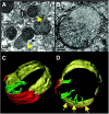
Mitochondrial herniation. (A) Electron micrograph of rat liver, 90 min after FAS activation. Arrow points to a herniation site, a large inner-membrane bleb protruding through a ruptured outer membrane. (B) A slice from an electron tomogram of a herniated mitochondrion. (C,D) Surface-rendered views showing the outer membrane (red), peripheral inner membrane (yellow), and cristae (green). Arrows point to crista junctions. Reproduced from Mootha et al. (2001) with permission (John Wiley and Sons, Inc.).

Similar articles

Cited by

References

    1. Alam Shibly S. U., Ghatak C., Sayem Karal M. A., Moniruzzaman M., Yamazaki M. (2016). Experimental estimation of membrane tension induced by osmotic pressure. Biophys. J. 111 2190–2201. 10.1016/j.bpj.2016.09.043 - DOI - PMC - PubMed
    1. Alexander C., Votruba M., Pesch U. E., Thiselton D. L., Mayer S., Moore A., et al. (2000). OPA1, encoding a dynamin-related GTPase, is mutated in autosomal dominant optic atrophy linked to chromosome 3q28. Nat. Genet. 26 211–215. 10.1038/79944 - DOI - PubMed
    1. Bay D. C., Hafez M., Young M. J., Court D. A. (2012). Phylogenetic and coevolutionary analysis of the beta-barrel protein family comprised of mitochondrial porin (VDAC) and Tom40. Biochim. Biophys. Acta 1818 1502–1519. 10.1016/j.bbamem.2011.11.027 - DOI - PubMed
    1. Blum T. B., Hahn A., Meier T., Davies K. M., Kuhlbrandt W. (2019). Dimers of mitochondrial ATP synthase induce membrane curvature and self-assemble into rows. Proc. Natl. Acad. Sci. U.S.A. 116 4250–4255. 10.1073/pnas.1816556116 - DOI - PMC - PubMed
    1. Chvanov M. (2006). Metabolic control of elastic properties of the inner mitochondrial membrane. J. Phys. Chem. B 110 22903–22909. 10.1021/jp0638181 - DOI - PubMed

LinkOut - more resources