. 2020 Jun 5:11:671.
doi: 10.3389/fphys.2020.00671. eCollection 2020.

Disentangling the Hypothesis of Host Dysosmia and SARS-CoV-2: The Bait Symptom That Hides Neglected Neurophysiological Routes

Affiliations

Disentangling the Hypothesis of Host Dysosmia and SARS-CoV-2: The Bait Symptom That Hides Neglected Neurophysiological Routes

Matteo Briguglio et al. Front Physiol. .

Abstract

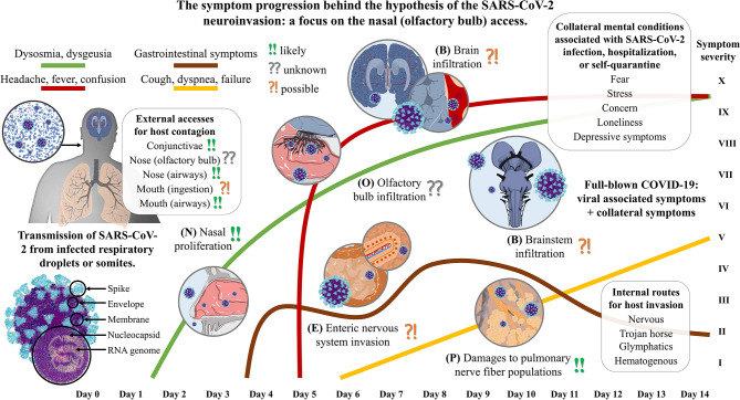
The respiratory condition COVID-19 arises in a human host upon the infection with SARS-CoV-2, a coronavirus that was first acknowledged in Wuhan, China, at the end of December 2019 after its outbreak of viral pneumonia. The full-blown COVID-19 can lead, in susceptible individuals, to premature death because of the massive viral proliferation, hypoxia, misdirected host immunoresponse, microthrombosis, and drug toxicities. Alike other coronaviruses, SARS-CoV-2 has a neuroinvasive potential, which may be associated with early neurological symptoms. In the past, the nervous tissue of patients infected with other coronaviruses was shown to be heavily infiltrated. Patients with SARS-CoV-2 commonly report dysosmia, which has been related to the viral access in the olfactory bulb. However, this early symptom may reflect the nasal proliferation that should not be confused with the viral access in the central nervous system of the host, which can instead be allowed by means of other routes for spreading in most of the neuroanatomical districts. Axonal, trans-synaptic, perineural, blood, lymphatic, or Trojan routes can gain the virus multiples accesses from peripheral neuronal networks, thus ultimately invading the brain and brainstem. The death upon respiratory failure may be also associated with the local inflammation- and thrombi-derived damages to the respiratory reflexes in both the lung neuronal network and brainstem center. Beyond the infection-associated neurological symptoms, long-term neuropsychiatric consequences that could occur months after the host recovery are not to be excluded. While our article does not attempt to fully comprehend all accesses for host neuroinvasion, we aim at stimulating researchers and clinicians to fully consider the neuroinvasive potential of SARS-CoV-2, which is likely to affect the peripheral nervous system targets first, such as the enteric and pulmonary nervous networks. This acknowledgment may shed some light on the disease understanding further guiding public health preventive efforts and medical therapies to fight the pandemic that directly or indirectly affects healthy isolated individuals, quarantined subjects, sick hospitalized, and healthcare workers.

Keywords: COVID-19; SARS-CoV-2; coronavirus; host pathogen interactions; infections; olfactory bulb; smell; virulence.


Figures

Figure 1
Figure 1
The symptom progression during the invasion of the human host by the SARS-CoV-2 upon nasal (olfactory bulb) access. The severe acute respiratory syndrome coronavirus that was discovered in Hubei province, China at the end of December 2019 (SARS-CoV-2) is a single-strand positive-sense RNA virus with the encoding potential of four structural proteins: the spike, the envelope, the membrane, and the nucleocapsid. The human host presents many potential accesses, but the inhalation of infected respiratory droplets exposes the airways first. (N) Assuming a nasal access, the olfactory mucosa may represent the primary site of the host for viral proliferation. It is reasonable to believe that SARS-CoV-2 needs to replicate several times before it can reach a harmful potential to infiltrate in the olfactory bulb, with the smell alterations being reported as one of the earliest symptoms. (O) The terminal nerve fibers extend in the olfactory epithelium colonized by the coronavirus. Here, not only axons of the fila olfactoria can be used as routes for central access, but also the trigeminal branches, the terminal nerve, blood or lymphatic vessels, or the cleft under the ensheathing cells of olfactory axons. (P) Upon inhalation, alveoli get in close contact with the virus, and it is reasonable to assume that the local inflammation could affect the different nerve fiber populations comprising the neuroendocrine cells and the annexed vagal contacts. Lung infection, which might manifest even few days after the contagion, can therefore provide direct access to the solitary tract and other parts of the central nervous system. (E) Once the virus has been accessed the epithelial barrier of gut cells through mucus or other vector ingestion, it can colonize the intestines that are permeated with the lymphatic system that drains the villi, the blood capillaries, and both the extrinsic and intrinsic neural networks. A fluctuating but chronic symptomatic onset is easily found. (B) It is still not clear which is the main route for the central nervous system access that may have a critical role in SARS-CoV-2 nervous infiltration, but both the brain and brainstem are likely to be infiltrated. High temperature is one of the earliest symptom after infection when more robust immune response is triggered, thus subsequently being associated with a permeabilization of the blood-brain barrier and immune cell infiltration. Cardiorespiratory centers in the medulla are critical districts that, if affected by either direct viral damages or indirect neuroinflammation and hypoxia, can lead to irreversible damage and fatal outcome.

Similar articles

Cited by

References

    1. Adriaensen D., Brouns I., Van Genechten J., Timmermans J. P. (2003). Functional morphology of pulmonary neuroepithelial bodies: extremely complex airway receptors. Anat. Rec. A Discov. Mol. Cell. Evol. Biol. 270, 25–40. 10.1002/ar.a.10007 - DOI - PubMed
    1. Akerlund A., Bende M., Murphy C. (1995). Olfactory threshold and nasal mucosal changes in experimentally induced common cold. Acta Otolaryngol. 115, 88–92. 10.3109/00016489509133353 - DOI - PubMed
    1. Bai Y., Yao L., Wei T., Tian F., Jin D. Y., Chen L., et al. (2020). Presumed asymptomatic carrier transmission of COVID-19. JAMA 323, 1406–1407. 10.1001/jama.2020.2565 - DOI - PMC - PubMed
    1. Baig A. M., Khaleeq A., Ali U., Syeda H. (2020). Evidence of the COVID-19 virus targeting the CNS: tissue distribution, host-virus interaction, and proposed neurotropic mechanisms. ACS Chem. Neurosci. 11, 995–998. 10.1021/acschemneuro.0c00122 - DOI - PubMed
    1. Bangash M. N., Patel J., Parekh D. (2020). COVID-19 and the liver: little cause for concern. Lancet Gastroenterol. Hepatol. 5:529–530. 10.1016/S2468-1253(20)30084-4 - DOI - PMC - PubMed