. 2020 Jun 3:11:893.
doi: 10.3389/fmicb.2020.00893. eCollection 2020.

Cadmium and Copper Cross-Tolerance. Cu+ Alleviates Cd2 + Toxicity, and Both Cations Target Heme and Chlorophyll Biosynthesis Pathway in Rubrivivax gelatinosus

Affiliations

Cadmium and Copper Cross-Tolerance. Cu+ Alleviates Cd2 + Toxicity, and Both Cations Target Heme and Chlorophyll Biosynthesis Pathway in Rubrivivax gelatinosus

Anne Soisig Steunou et al. Front Microbiol. .

Abstract

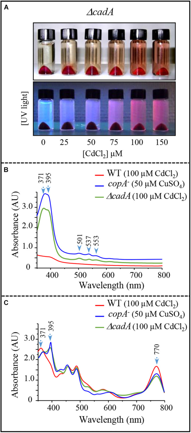
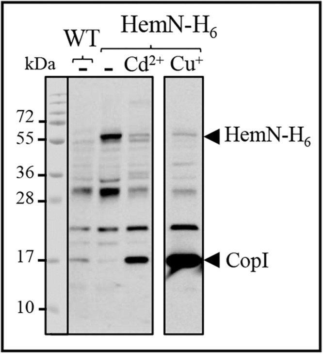
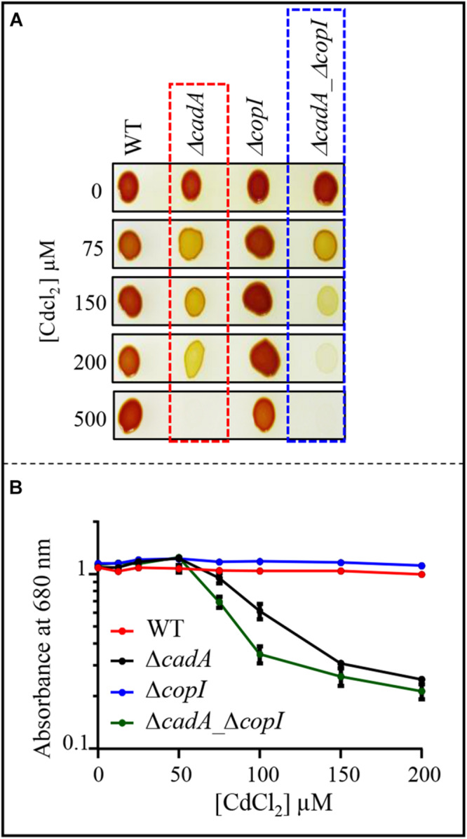
Cadmium, although not redox active is highly toxic. Yet, the underlying mechanisms driving toxicity are still to be characterized. In this study, we took advantage of the purple bacterium Rubrivivax gelatinosus strain with defective Cd2 +-efflux system to identify targets of this metal. Exposure of the ΔcadA strain to Cd2 + causes a decrease in the photosystem amount and in the activity of respiratory complexes. As in case of Cu+ toxicity, the data indicated that Cd2 + targets the porphyrin biosynthesis pathway at the level of HemN, a S-adenosylmethionine and CxxxCxxC coordinated [4Fe-4S] containing enzyme. Cd2 + exposure therefore results in a deficiency in heme and chlorophyll dependent proteins and metabolic pathways. Given the importance of porphyrin biosynthesis, HemN represents a key metal target to account for toxicity. In the environment, microorganisms are exposed to mixture of metals. Nevertheless, the biological effects of such mixtures, and the toxicity mechanisms remain poorly addressed. To highlight a potential cross-talk between Cd2 + and Cu+ -efflux systems, we show (i) that Cd2 + induces the expression of the Cd2 +-efflux pump CadA and the Cu+ detoxification system CopA and CopI; and (ii) that Cu+ ions improve tolerance towards Cd2 +, demonstrating thus that metal mixtures could also represent a selective advantage in the environment.

Keywords: CadA/ZntA; [4Fe-4S]; cadmium/copper; cross-talk; metal homeostasis; metal toxicity; porphyrin biosynthesis.


Figures

FIGURE 1
FIGURE 1
(A) Genetic organization of the cadAR locus in R. gelatinosus. To inactivate cadA, the NarI fragment was replaced by antibiotic resistance Tp cassette. (B) Growth phenotype of the wild-type (WT) and ΔcadA on solid medium supplemented with CdCl2. Plates were incubated under aerobic respiration (RES) in the dark or under photosynthesis (PS) for 24h at 30°C prior to photography. (C,D) Growth inhibition of the WT and ΔcadA mutant challenged with increasing CdCl2 concentrations under photosynthesis (PS) (C) and respiration (RES) (D) conditions. Cells were grown for 24 h at 30°C before OD680nm measurement. The error bars represent the standard deviation of the mean of 3 independent experiments.
FIGURE 2
FIGURE 2
Expression profile of CadA and CopI in R. gelatinosus wild-type (WT) and ΔcadA cells challenged with Cd2 +. Cells were grown overnight (18 h) under photosynthesis and total protein extract from the same amount of cells (OD680nm = 0.1) were separated on a 15% Tris-glycine SDS-PAGE (A). Periplasmic fractions were purified from ΔcadA and ΔcadA_ΔcopI cells challenged or not with Cd2 + and separated on a 15% Tris-glycine SDS-PAGE (B). Proteins were visualized after Western blotting using the HRP-HisProbe (Pierce).
FIGURE 3
FIGURE 3
Expression level of CadA and CopI in R. gelatinosus wild-type (WT) and cadR- mutant cells challenged with increasing concentration of CdCl2. Cells were grown overnight (18 h) under photosynthetic condition. Total protein extract from the same amount of cells (OD680nm = 0.1) were separated on a 15% Tris-glycine SDS-PAGE. Proteins were visualized after Western blotting using the HRP-HisProbe.
FIGURE 4
FIGURE 4
Cadmium effect on photosynthesis and respiratory membrane complexes in the wild-type (WT) and ΔcadA mutant grown overnight (18 h) under microaerobic condition. (A) Effect of CdCl2 on photosystem (860–800 nm) amount in the membranes. (B) Difference (reduced minus oxidized) pyridine haemochrome spectra of WT (red) and ΔcadA (black) membranes with 100 μM (dashed lines) or without cadmium (lines). (C) cbb3 cytochrome c oxidase and succinate dehydrogenase (SDH) in-gel activity assays. Equal amount of DDM-solubilized membrane proteins from wild-type (WT) and ΔcadA cells grown without or in the presence of 150 μM CdCl2 were separated on 3–12% acrylamide-bisacrylamide gradient BN-PAGE. Gels were first assayed for cbb3 cytochrome c oxidase activity (DAB staining) and subsequently assayed for SDH activity (Succinate/NBT). The photosynthetic RC-LH complexes are also visible on the gel thanks to their photopigments.
FIGURE 5
FIGURE 5
Cadmium effect on total heme (550 nm) and bacteriochlorophyll (756 nm) content in the wild-type (WT) (A) and ΔcadA strain (B) grown by photosynthesis for 18 h in the presence of CdCl2.
FIGURE 6
FIGURE 6
Effect of cadmium on photosynthetic growth in the ΔcadA strain and characterization of the pigment extruded in the culture medium. (A) Phenotype of the ΔcadA mutant grown in the presence of increasing CdCl2 concentration. Appearance of coproporphyrin III in the spent medium under visible and UV light. (B) UV-visible absorption spectra of the spent medium from the wild-type and ΔcadA strain grown with 100 μM CdCl2, in comparison with the spent medium of copA strain that accumulates coproporphyrin III (395–371 nm) when grown in the presence of 50 μM CuSO4. (C) Absorption spectra of total pigment extract from membranes of the wild-type, ΔcadA cells grown with 100 μM CdCl2 and copA cells grown with 50 μM CuSO4. The spectra show the decrease in the amount of bacteriochlorophyll a (770 nm) in ΔcadA and copA mutants and reveal the presence of coproporphyrin III in these cells.
FIGURE 7
FIGURE 7
Expression level of HemN-H6 in R. gelatinosus WT and HemN-H6 strain in malate medium (-) or challenged with 1000 μM CdCl2 (Cd2 +) or 500 μM CuSO4 (Cu+). Cells were grown overnight (18 h) under photosynthetic condition. Soluble protein fractions (20 μg) were separated on a 15% Tris-glycine SDS-PAGE. Proteins were visualized after Western blotting using the HRP-HisProbe.
FIGURE 8
FIGURE 8
(A) Growth phenotype of the wild-type (WT), ΔcadA, ΔcopI, and ΔcadAcopI mutants in the presence of increasing CdCl2 concentrations on solid plates. Cells were grown by photosynthesis for 48 h at 30°C prior to photography. (B) Growth inhibition of the wild-type, ΔcadA, ΔcopI, and ΔcadAcopI mutants challenged with increasing CdCl2 concentration. Cells were grown in liquid under photosynthetic condition for 21 h at 30°C before OD680nm measurement. The error bars represent the standard deviation of the mean of 3 independent experiments.
FIGURE 9
FIGURE 9
Effect of CuSO4 on growth in the presence of increasing concentration of CdCl2. (A) Growth phenotype of the wild-type (WT), ΔcadA and copA strains in presence of increasing CdCl2 concentration with or without addition of 200 μM CuSO4 on solid plates. (B) Growth inhibition of the wild-type and ΔcadA mutant challenged with increasing CdCl2 concentration in the medium supplemented or not with 200 μM CuSO4. Cells were grown under photosynthetic condition for 18 h at 30°C before OD680nm measurement. The error bars represent the standard deviation of the mean of 3 independent experiments. (C) Expression level of CopA-H6 and CopI in R. gelatinosus ΔcadA-copA-H6 mutant challenged with increasing concentration of CuSO4, CdCl2, or CdCl2 + CuSO4. Cells were grown in the malate (M) medium supplemented or not with metals, under photosynthetic condition. Total protein extract from the same amount of cells (OD680nm = 0.1) were separated on a 15% tris-glycine SDS-PAGE. Proteins were visualized after Western blotting using the HRP-HisProbe.

References

    1. Arguello J. M., Eren E., Gonzalez-Guerrero M. (2007). The structure and function of heavy metal transport P1B-ATPases. Biometals 20 233–248. 10.1007/s10534-006-9055-6 - DOI - PubMed
    1. Azzouzi A., Steunou A. S., Durand A., Khalfaoui-Hassani B., Bourbon M. L., Astier C., et al. (2013). Coproporphyrin III excretion identifies the anaerobic coproporphyrinogen III oxidase HemN as a copper target in the Cu+-ATPase mutant copA– of Rubrivivax gelatinosus. Mol. Microbiol. 88 339–351. 10.1111/mmi.12188 - DOI - PubMed
    1. Baekgaard L., Mikkelsen M. D., Sorensen D. M., Hegelund J. N., Persson D. P., Mills R. F., et al. (2010). A combined zinc/cadmium sensor and zinc/cadmium export regulator in a heavy metal pump. J. Biol. Chem. 285 31243–31252. 10.1074/jbc.m110.111260 - DOI - PMC - PubMed
    1. Barwinska-Sendra A., Waldron K. J. (2017). The Role of Intermetal Competition and Mis-Metalation in Metal Toxicity. Adv. Microb. Physiol. 70 315–379. 10.1016/bs.ampbs.2017.01.003 - DOI - PubMed
    1. Breckau D., Mahlitz E., Sauerwald A., Layer G., Jahn D. (2003). Oxygen-dependent coproporphyrinogen III oxidase (HemF) from Escherichia coli is stimulated by manganese. J. Biol. Chem. 278 46625–46631. 10.1074/jbc.m308553200 - DOI - PubMed