. 2020 Jun 4:11:1100.
doi: 10.3389/fmicb.2020.01100. eCollection 2020.

Characterization of a Novel Functional Trimeric Catechol 1,2-Dioxygenase From a Pseudomonas stutzeri Isolated From the Gulf of Mexico

Affiliations

Characterization of a Novel Functional Trimeric Catechol 1,2-Dioxygenase From a Pseudomonas stutzeri Isolated From the Gulf of Mexico

Julieta Rodríguez-Salazar et al. Front Microbiol. .

Abstract

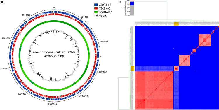
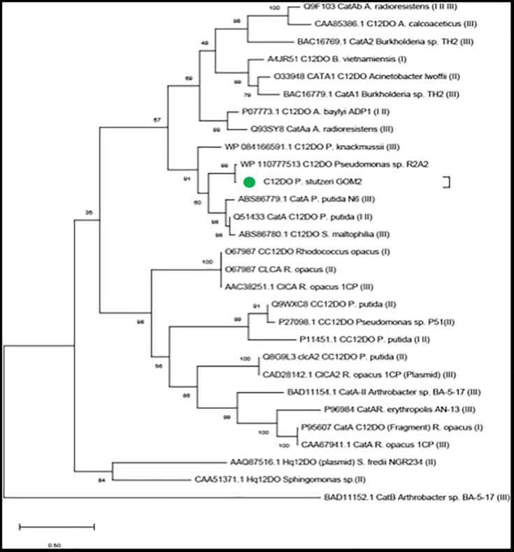
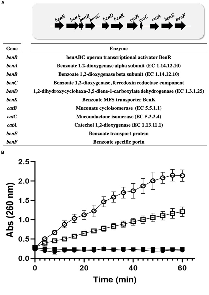
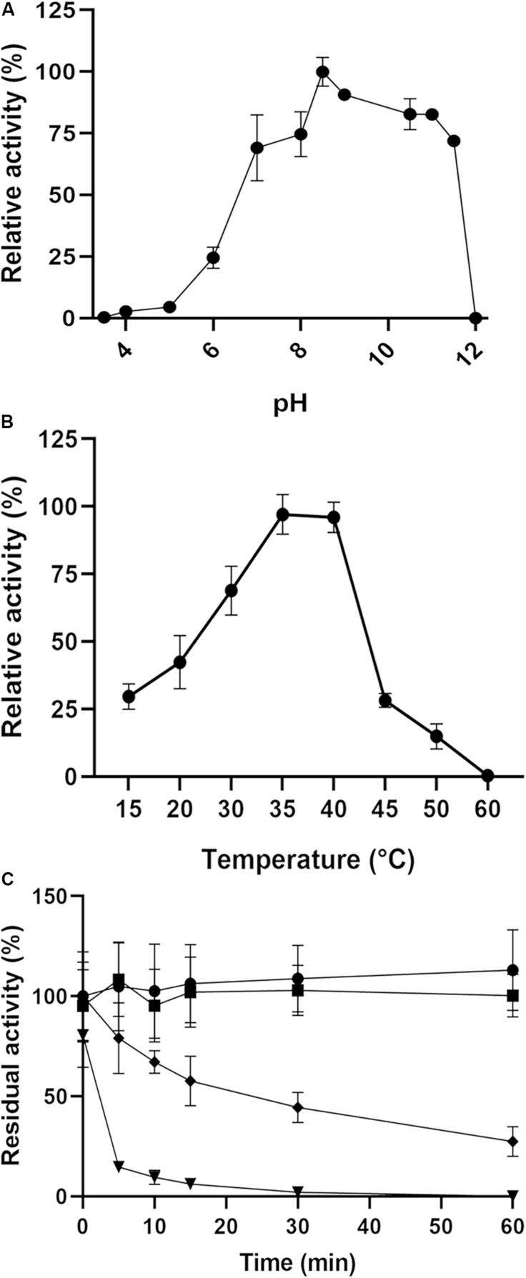
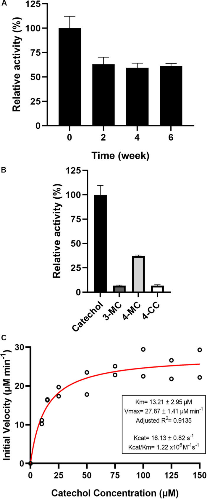
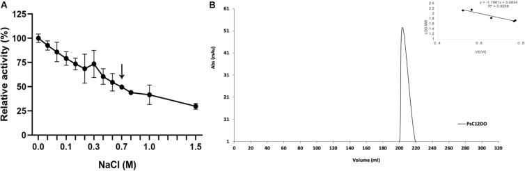
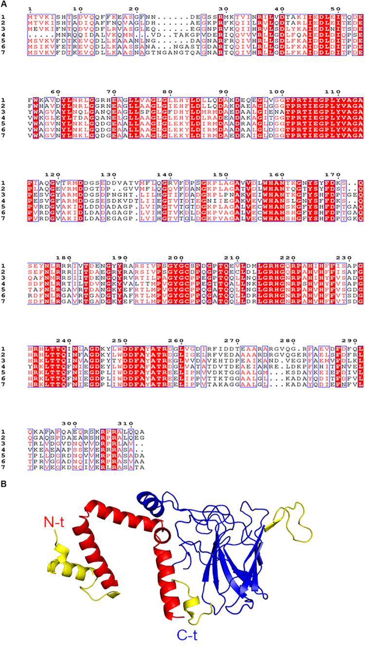
Catechol 1,2 dioxygenases (C12DOs) have been studied for its ability to cleavage the benzene ring of catechol, the main intermediate in the degradation of aromatic compounds derived from aerobic degradation of hydrocarbons. Here we report the genome sequence of the marine bacterium Pseudomonas stutzeri GOM2, isolated from the southwestern Gulf of Mexico, and the biochemical characterization of its C12DO (PsC12DO). The catA gene, encoding PsC12DO of 312 amino acid residues, was cloned and expressed in Escherichia coli. Many C12DOs have been described as dimeric enzymes including those present in Pseudomonas species. The purified PsC12DO enzyme was found as an active trimer, with a molecular mass of 107 kDa. Increasing NaCl concentration in the enzyme reaction gradually reduced activity; in high salt concentrations (0.7 M NaCl) quaternary structural analysis determined that the enzyme changes to a dimeric arrangement and causes a 51% decrease in specific activity on catechol substrate. In comparison with other C12DOs, our enzyme showed a broad range of action for PsC12DO in solutions with pH values ranging from neutral to alkaline (70%). The enzyme is still active after incubation at 50°C for 30 min and in low temperatures to long term storage after 6 weeks at 4°C (61%). EDTA or Ca2+ inhibitors cause no drastic changes on residual activity; nevertheless, the activity of the enzyme was affected by metal ions Fe3+, Zn2+ and was completely inhibited by Hg2+. Under optimal conditions the k cat and K m values were 16.13 s-1 and 13.2 μM, respectively. To our knowledge, this is the first report describing the characterization of a marine C12DOs from P. stutzeri isolated from the Gulf of Mexico that is active in a trimeric state. We consider that our enzyme has important features to be used in environments in presence of EDTA, metals and salinity conditions.

Keywords: Pseudomonas; aromatic compounds; catechol degradation; intradiol dioxygenase; trimeric structure.


Figures

FIGURE 1
FIGURE 1
Genome assembly and ANI comparison. (A) Circular diagram of P. stutzeri GOM2 showing (from inner to outer)% G + C, GC skew and scaffolds. (B) Average nucleotide identity heatmap with all the available P. stutzeri genomes from GenBank. The yellow box indicates the group to which P. stutzeri GOM2 is related.
FIGURE 2
FIGURE 2
Evolutionary analyses. The green circle denotes the sequence of P. stutzeri GOM2. In the parentheses the reference is indicated where the protein sequence was obtained: (I) Nazmi et al. (2019), (II) Caglio et al. (2009), and (III) Guzik et al. (2013).
FIGURE 3
FIGURE 3
Induction of ortho pathway in Pseudomonas. (A) Benzoate catabolism operon in P. stutzeri GOM2. (B) Cis,cis-muconate production by Pseudomonas strain crude extracts. P. stutzeri GOM2 grown with BS (closed circles) or BA (open circles) and P. putida KT2440 grown with BS (closed squares) or BA (open squares) are shown. The assay was carried out in 96-well microplates, using 10 μg/mL total crude protein, with a 50 mM catechol solution in PBS buffer, pH 7.4, at 30°C. Values are the averages of three measurements, and bars indicate the SD values.
FIGURE 4
FIGURE 4
Size exclusion chromatography of PsC12DO. (A) SDS–PAGE analysis of purified PsC12DO. MW, molecular weight ladder. Lane 1, crude extract. Lane 2, crude extract after the addition of IPTG. Lane 3, purified PsC12DO. (B) Chromatogram showing the elution profile of PsC12DO at 180 mL (108 kDa). On the right, a five-point calibration graph.
FIGURE 5
FIGURE 5
Effects of pH and temperature on the enzyme activity of PsC12DO. (A) Activity assays were performed in 50 mM sodium acetate (pH 3.6–5.0), 50 mM Sorensen (pH 6.0–8.5), and 50 mM glycine-NaOH (pH 8.5–12) at 40°C. (B) Activity assays were performed in a range of 15–60°C to determine the optimal temperature condition. (C) Thermal stability assays were performed in a range of 40–60°C. Temperatures of 40°C (circles), 45°C (squares), 50°C (diamonds) 60°C (triangles) in 50 mM glycine-NaOH buffer pH 8.5. Values are the average of three measurements, and bars indicate the SD values.
FIGURE 6
FIGURE 6
(A) Storage stability of PsC12DO at 4°C. (B) Substrate specificity of PsC12DO. Catechol (black), 3-MC (gray), 4-MC (light gray), and 4-CC (white) are shown. (C) Effect of catechol concentration on enzyme activity and kinetic parameters. Values are the averages of three measurements, and bars indicate the SD values.
FIGURE 7
FIGURE 7
Effects of NaCl on the enzyme activity of PsC12DO. (A) Activity assays were performed in glycine-NaOH buffer, pH 8.5, supplemented with NaCl at 40°C. Values are the average of three measurements, and bars indicate the SD values. (B) Chromatogram showing the elution profile of PsC12DO at 203 mL (89 kDa) in 50 mM glycine-NaOH, pH 8.5, supplemented with 700 mM NaCl. On the right, a five-point calibration graph.
FIGURE 8
FIGURE 8
(A) Amino acid sequence alignment of six catechol 1,2-dioxygenase homologs to PSC12DO. Residues in red are conserved in all sequences. Residues in a blue frame have similarity. (1) Catechol 1,2-dioxygenase Pseudomonas stutzeri (PsC12DO), (2) Catechol 1,2-dioxygenase Pseudomonas Arvilla (PDBid:2AZQ), (3) Catechol 1,2-dioxygenase Acinetobacter calcoaceticus (PDBid:1DLM), (4) Catechol 1,2-dioxygenase Acinetobacter radioresistens (PDBid:2XSR), (5) Catechol 1,2-dioxygenase Burkholderia multivorans (PDBid:5UMH), (6) Catechol 1,2-dioxygenase Burkholderia vietnamiensis (PDBid:5TD3), (7) Catechol 1,2-dioxygenase Burkholderia ambifaria (PDBid:5VXT). The alignment was generated by using Clustal Omega. (B) A model of monomeric PsC12DO generated by CHPModel 3.2 using the structure PDBid:2AZQ as reference.

Similar articles

Cited by

References

    1. Al-Hakim M. H., Hasan R., Ali M. H., Rabbee M. F., Marufatuzzahan H. M., Joy Z. F. (2015). In-silico characterization and homology modeling of catechol 1,2 dioxygenase involved in processing of catechol- an intermediate of aromatic compound degradation pathway. Glob. J. Sci. Front. Res. G Bio-Tech Genet. 15 1–13.
    1. Ashton A. R., Siegel G. M. (1997). Stimulation of spinach (Spinacia oleracea) chloroplast fructose-1,6-bisphosphatase by mercuric ions. FEBS Lett. 408 30–32. 10.1016/s0014-5793(97)00385-2 - DOI - PubMed
    1. Atlas R. M. (1981). Microbial degradation of petroleum hydrocarbons: an environmental perspective. Microbiol. Rev. 45 180–209. - PMC - PubMed
    1. Bagdasarian M., Lurz R., Ruckert B., Franklin F. C., Bagdasarian M. M., Frey J., et al. (1981). Specific-purpose plasmid cloning vectors. II. Broad host range, high copy number, RSF1010-derived vectors, and a host-vector system for gene cloning in Pseudomonas. Gene 16 237–247. 10.1016/0378-1119(81)90080-9 - DOI - PubMed
    1. Bonomo R., Cennamo G., Purrello R., Santoro A., Zappala R. (2001). Comparison of three fungal laccases from Rigidoporus lignosus and Pleurotus ostreatus: correlation between conformation changes and catalytic activity. J. Inorg. Biochem. 83 67–75. 10.1016/s0162-0134(00)00130-6 - DOI - PubMed

LinkOut - more resources