Review
. 2020 Jun 2:11:1066.
doi: 10.3389/fimmu.2020.01066. eCollection 2020.

Phagocytosis: Our Current Understanding of a Universal Biological Process

Affiliations
Review

Phagocytosis: Our Current Understanding of a Universal Biological Process

Eileen Uribe-Querol et al. Front Immunol. .

Abstract

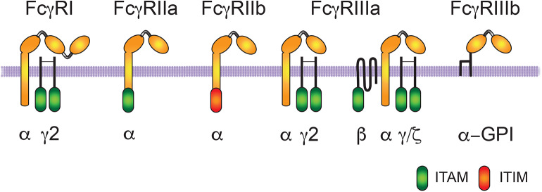
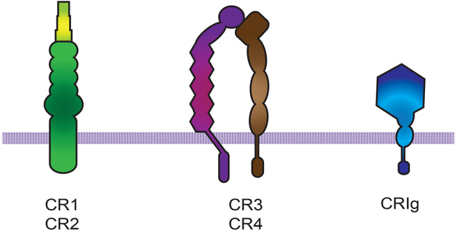
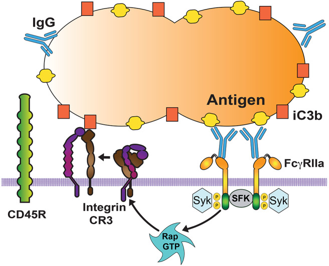
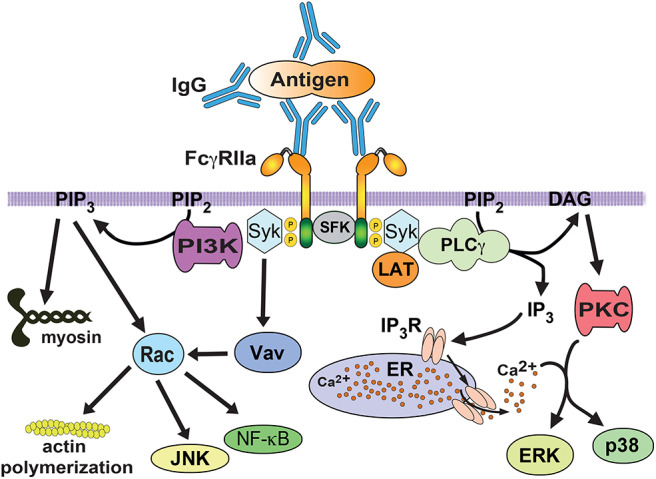
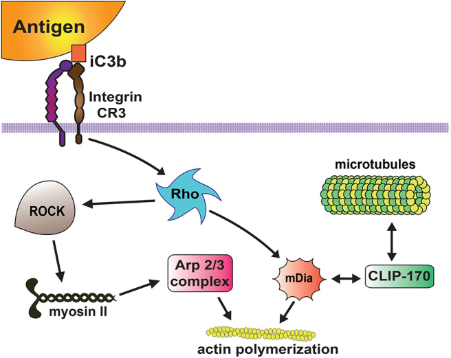
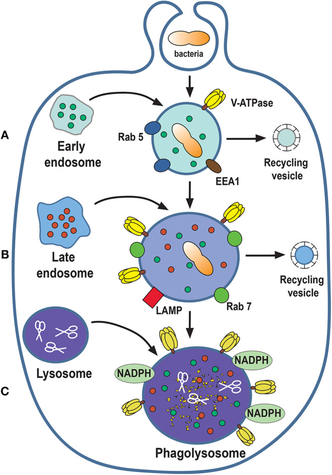
Phagocytosis is a cellular process for ingesting and eliminating particles larger than 0.5 μm in diameter, including microorganisms, foreign substances, and apoptotic cells. Phagocytosis is found in many types of cells and it is, in consequence an essential process for tissue homeostasis. However, only specialized cells termed professional phagocytes accomplish phagocytosis with high efficiency. Macrophages, neutrophils, monocytes, dendritic cells, and osteoclasts are among these dedicated cells. These professional phagocytes express several phagocytic receptors that activate signaling pathways resulting in phagocytosis. The process of phagocytosis involves several phases: i) detection of the particle to be ingested, ii) activation of the internalization process, iii) formation of a specialized vacuole called phagosome, and iv) maturation of the phagosome to transform it into a phagolysosome. In this review, we present a general view of our current understanding on cells, phagocytic receptors and phases involved in phagocytosis.

Keywords: ERK; antibody; complement; immunoglobulin; integrin; neutrophil; phagocytosis.


Figures

Figure 1
Figure 1
Human Fcγ receptors. The human receptors for the Fc portion of immunoglobulin G (IgG) molecules are classified in three groups FcγRI, FcγRII, and FcγRIII. The IgG binding α-subunit in the high affinity FcγRI, possesses three immunoglobulin (Ig)-like extracellular domains. The α-subunit in the other low-affinity receptors presents only two Ig-like domains. Activating receptors contain an ITAM (immunoreceptor tyrosine-based activation motif) sequence within the α subunit (for FcγRIIa) or within the accessory γ and ζ chains (for FcγRI and FcγRIIIa). FcγRIIIa has a homodimer of γ chains in macrophages, natural killer (NK) cells, and dendritic cells, whereas it has a heterodimer of γ/ζ chains and an extra β chain in basophils and mast cells. FcγRIIIb is also an activating receptor, which is bound to the cell membrane via a glycosylphosphatidylinositol (GPI) anchor. In contrast, FcγRIIb is an inhibitory receptor containing an ITIM (immunoreceptor tyrosine-based inhibition motif) sequence.
Figure 2
Figure 2
Complement receptors. There are three groups of complement receptors: (i) the short consensus repeat (SCR) modules that code for CR1 and CR2, (ii) the β2 integrin family members CR3 and CR4 (66), and (iii) the immunoglobulin Ig-superfamily member CRIg.
Figure 3
Figure 3
Cooperation among phagocytic receptors. Most phagocytic receptors, such as receptors for antibody (FcγRIIa) and receptors for complement (Integrin CR3) cooperate to bind the particle to be ingested. FcγR aggregation triggers an inside-out signal that activates integrins via the GTPase Rap. Activated Rap (Rap GTP) is responsible for integrin activation. Then, activated integrins also bind to the particle (via complement fragment C3b), and form a diffusion barrier that excludes larger molecules, such as the transmembrane phosphatase CD45. This allows other Fc receptors to be engaged and increase the signaling for phagocytosis. SFK, Src family kinases. Syk, spleen tyrosine kinase.
Figure 4
Figure 4
FcγR signaling for phagocytosis. FcγRIIa crosslinking by immunoglobulin (IgG) bound to a particle, induces activation of Src family kinases (SFK), which phosphorylate tyrosine residues in the ITAM sequence within the cytoplasmic tail of the receptor. Then, spleen tyrosine kinase (Syk) associates with phosphorylated ITAMs and leads to phosphorylation and activation of a signaling complex formed by the scaffold protein LAT (linker for activation of T cells) interacting with various proteins. One of these proteins is phospholipase C gamma (PLCγ), which produces inositoltrisphosphate (IP3), and diacylglycerol (DAG). These second messengers cause calcium release and activation of protein kinase C (PKC), respectively. PKC leads to activation of extracellular signal-regulated kinases (ERK and p38). The guanine nucleotide exchange factor Vav activates the GTPase Rac, which is involved in regulation of the actin cytoskeleton. Rac is also involved in activation of transcription factors such as NF-κB and JNK. The enzyme phosphatidylinositol 3-kinase (PI3K), which is recruited and activated by Syk, generates the lipid phosphatidylinositol-3,4,5-trisphosphate (PIP3) at the phagocytic cup. This lipid also regulates Rac activation, and contractile proteins such as myosin. P represents a phosphate group. ER, endoplasmic reticulum. IP3R, receptor (calcium channel) for inositoltrisphosphate.
Figure 5
Figure 5
Complement receptor signaling for phagocytosis. The complement receptor 3 (CR3 integrin) binds the complement molecules (iC3b) deposited on the target particle, and activates a signaling pathway that leads to activation of the GTPase Rho. Then, active Rho induces actin polymerization via two mechanisms. Rho activates Rho kinase (ROCK), which phosphorylates and activates myosin II, inducing accumulation of Arp2/3 and actin assembly at the phagocytic cup. Rho also promotes accumulation of mDia1 (mammalian diaphanous-related formin 1), which stimulates linear actin polymerization. In addition, mDia1 binds directly to the microtubule-associated protein CLIP-170 providing a link to the microtubule cytoskeleton.
Figure 6
Figure 6
Phagosome maturation. The nascent phagosome gets transformed into a microbicidal vacuole, the phagolysosome, by sequential interactions with vesicles from the endocytic pathway. The process can be described in three stages of maturation: early (A), late (B), and phagolysosome (C). In this process, composition of the membrane changes to include molecules that control membrane fusion, such as the GTPases Rab5 and Rab7. The phagolysosome becomes increasingly acidic by the action of a proton-pumping V-ATPase and acquires various degradative enzymes, such as cathepsins, proteases, lysozymes, and lipases (scissors). EEA1, early endosome antigen 1; LAMP, lysosomal-associated membrane protein; NADPH, nicotinamide adenine dinucleotide phosphate oxidase.

References

    1. Rabinovitch M. Professional and non-professional phagocytes: an introduction. Trends Cell Biol. (1995) 5:85–7. 10.1016/S0962-8924(00)88955-2 - DOI - PubMed
    1. Gordon S. Phagocytosis: an immunobiologic process. Immunity. (2016) 44:463–75. 10.1016/j.immuni.2016.02.026 - DOI - PubMed
    1. Levin R, Grinstein S, Canton J. The life cycle of phagosomes: formation, maturation, and resolution. Immunol Rev. (2016) 273:156–79. 10.1111/imr.12439 - DOI - PubMed
    1. Canton J. Phagosome maturation in polarized macrophages. J Leukoc Biol. (2014) 96:729–38. 10.1189/jlb.1MR0114-021R - DOI - PubMed
    1. Dambuza IM, Brown GD. C-type lectins in immunity: recent developments. Curr Opin Immunol. (2015) 32:21–7. 10.1016/j.coi.2014.12.002 - DOI - PMC - PubMed

Publication types

MeSH terms

Substances