Review
. 2020 Jun 2:11:539.
doi: 10.3389/fgene.2020.00539. eCollection 2020.

Enzymatic Functions for Toll/Interleukin-1 Receptor Domain Proteins in the Plant Immune System

Affiliations
Review

Enzymatic Functions for Toll/Interleukin-1 Receptor Domain Proteins in the Plant Immune System

Adam M Bayless et al. Front Genet. .

Abstract

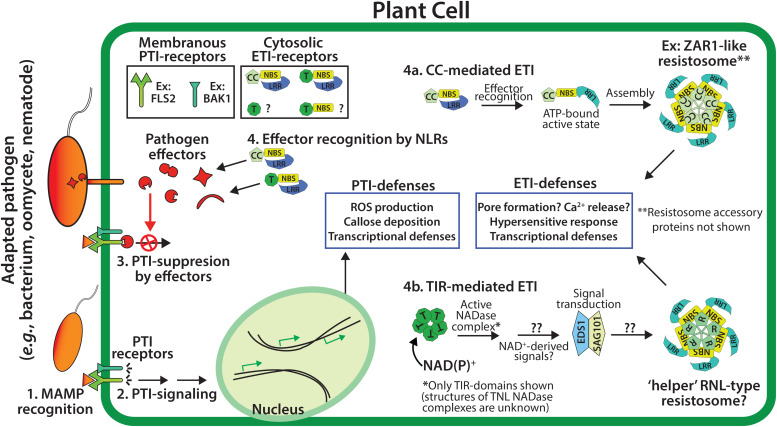
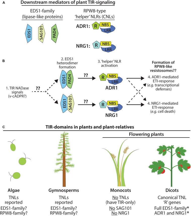
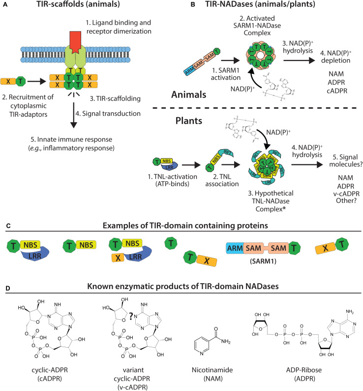
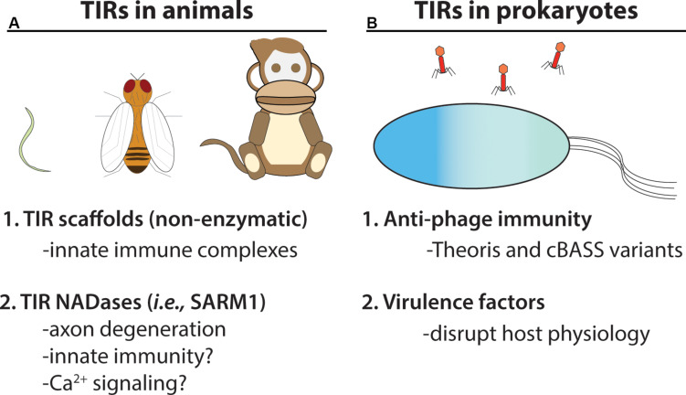
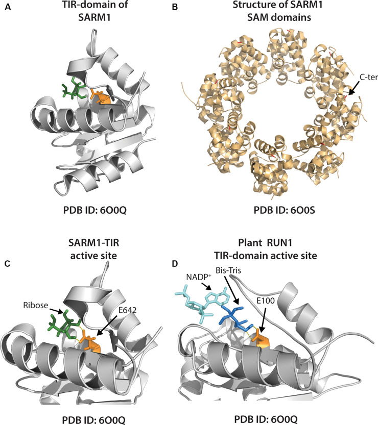
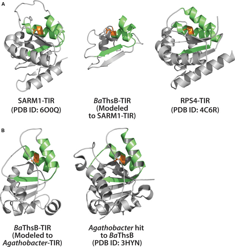
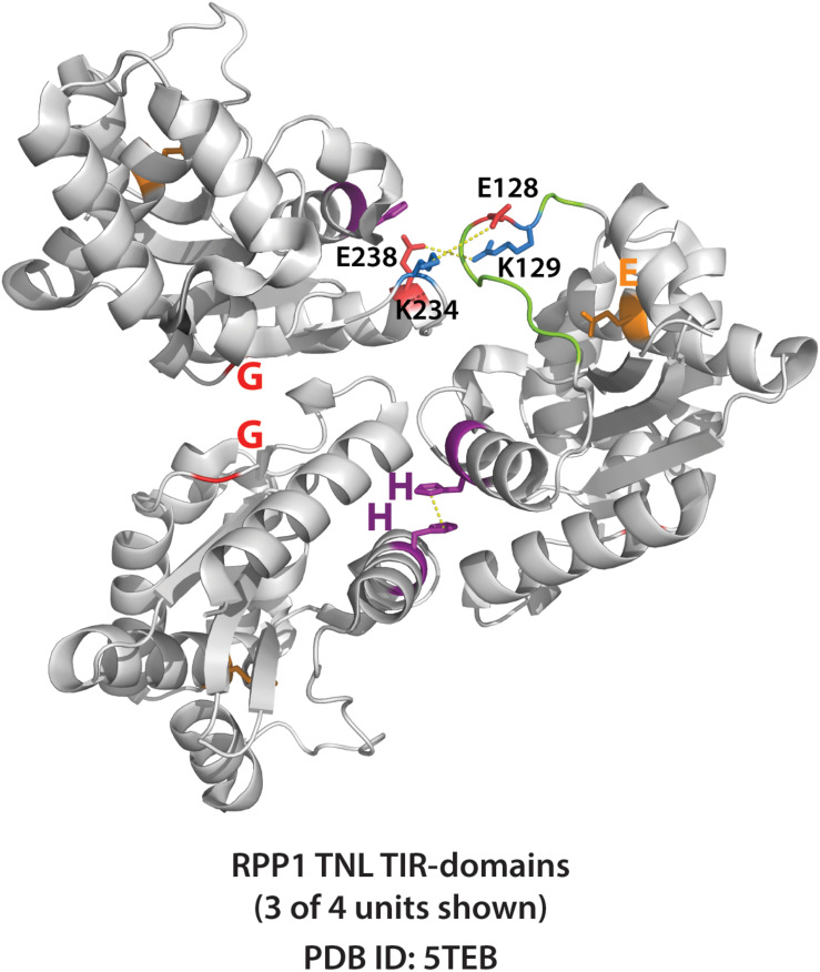
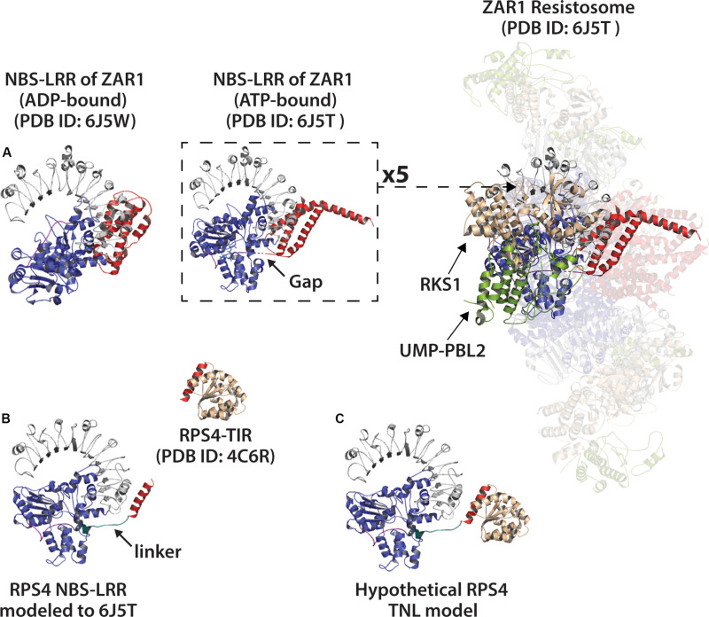
Rationally engineered improvements to crop plants will be needed to keep pace with increasing demands placed on agricultural systems by population growth and climate change. Engineering of plant immune systems provides an opportunity to increase yields by limiting losses to pathogens. Intracellular immune receptors are commonly used as agricultural disease resistance traits. Despite their importance, how intracellular immune receptors confer disease resistance is still unknown. One major class of immune receptors in dicots contains a Toll/Interleukin-1 Receptor (TIR) domain. The mechanisms of TIR-containing proteins during plant immunity have remained elusive. The TIR domain is an ancient module found in archaeal, bacterial and eukaryotic proteins. In animals, TIR domains serve a structural role by generating innate immune signaling complexes. The unusual animal TIR-protein, SARM1, was recently discovered to function instead as an enzyme that depletes cellular NAD+ (nicotinamide adenine dinucleotide) to trigger axonal cell death. Two recent reports have found that plant TIR proteins also have the ability to cleave NAD+. This presents a new paradigm from which to consider how plant TIR immune receptors function. Here, we will review recent reports of the structure and function of TIR-domain containing proteins. Intriguingly, it appears that TIR proteins in all kingdoms may use similar enzymatic mechanisms in a variety of cell death and immune pathways. We will also discuss TIR structure-function hypotheses in light of the recent publication of the ZAR1 resistosome structure. Finally, we will explore the evolutionary context of plant TIR-containing proteins and their downstream signaling components across phylogenies and the functional implications of these findings.

Keywords: NADase; NLR; TIR; Toll/interleukin-1 receptor; innate immunity.


Figures

FIGURE 1
FIGURE 1
Overview of the two tiers of the plant immune system. PTI (pattern-triggered immunity) utilizes membrane associated PTI receptors to detect conserved microbial associated molecular patterns (MAMPs, e.g., chitin or flagellin), and signal downstream PTI-immunity (steps 1 and 2). The effectors of adapted pathogens can disarm PTI-immunity (step 3), while genetically resistant plants utilize NLR-type (NBS-LRR) resistance proteins to detect effector activities and trigger ETI (effector-triggered immunity, step 4), which often includes localized cell death - the hypersensitive response (HR). Plant NLR resistance proteins generally possess N-terminal CC (coiled-coil) or TIR (Toll/interleukin-1 Receptor) domains.
FIGURE 2
FIGURE 2
Immune signaling by plant TIR NADases requires downstream components. (A) Members of the EDS1 lipase-like family: EDS1, SAG101, PAD4. Terminal mediators of TIR-signal transduction are the RPW8-type ‘helper’ RNLs: ADR1 and/or NRG1. (B) Model of ETI-pathway activation by plant TIR NADases. Perception of plant TIR signals (e.g., v-cADPR?) promotes EDS1-family heterodimerization, and subsequent activation of the ‘helper’ RNLs, ADR1 or NRG1. EDS1-PAD4 heterodimers may favor activation of ADR1-mediated responses (transcriptional defense programs), while EDS1-SAG101 heterodimers activate NRG1-mediated responses (cell death). Functional redundancy among NRG1 and ADR1 indicated by dashed arrows. (C) TIR-domain containing proteins, including TNLs, are found in the genomes of phylogenetically distant plant-lineages and in the relatives of land plants, including green algae (Sun et al., 2014; Shao et al., 2019), as well as gymnosperms (western white pine) and the moss, Physcomitrella patens. Monocots do not encode TNLs and lack two downstream mediators of TIR-immune signaling: SAG101 and NRG (Collier et al., 2011; Lapin et al., 2019).
FIGURE 3
FIGURE 3
Model of TIR-domain scaffolding (animals) and TIR-NADase activity (plants and animals). (A) Canonical TIR-scaffold function in animals: TIR-TIR interactions promote signal complex formation and innate immune signal transduction. (B) Top: animal TIR NADases (e.g., SARM1) assemble into high order complexes, and hydrolyze NAD(P)+ substrate and alter NAD(P)+ pools. Bottom: assembly of plant TIR-domains into hypothetical NADase complex (resistosome-like?) and generation of immunomodulatory signals. (C) Numerous TIR-domain configurations are present in animal, plant, and bacterial proteins. Plant TIR-domains are often found in modular NBS-LRRs, TIR-NBS, TIR-X or TIR-only proteins. -X corresponds to atypical or undefined domains. The animal SARM1 TIR is located at the C-terminus; the SARM1 SAM-domains promote oligomerization. (D) Known products of TIR NADases; plant TIRs produce variant cyclic-ADPR (v-cADPR), whose structure is currently unknown.
FIGURE 4
FIGURE 4
TIR NADases were recently reported in animals and prokaryotes. (A) Diverse eukaryotic organisms, including invertebrates (e.g., C. elegans, D. melanogaster) and vertebrates, utilize TIR-domain containing proteins in cellular innate immunity. Non-enzymatic TIR-domain containing proteins in animals promote signal complex formation via TIR – TIR interactions. The SARM1 NADase TIR from animals functions in axon degeneration, and is reported to function in immunity in C. elegans (Shivers et al., 2009). (B) Numerous bacteria and archaeal species encode TIR-NADases. Prokaryotic TIR-domain containing proteins are reported to function in anti-phage immunity (Thoeris system and variants of cBASS) (Doron et al., 2018; Cohen et al., 2019). TIR-domains from pathogenic bacteria are reported to function in virulence (Alaidarous et al., 2014).
FIGURE 5
FIGURE 5
Structures of individual animal and plant TIR-domain NADases, and the higher order SARM1 SAM octamer. (A) Crystal structure of the SARM1-TIR domain (PDB ID: 6O0Q) with ribose (shown green) positioned near putative catalytic glutamate residue (E642), colored orange. (B) Crystal structure of tandem SAM domains of the animal TIR-NADase, SARM1 (PDB ID: 6O0S). The SARM1 SAM domains adopt a closed octameric ring conformation. C-terminal end of SAM tipped with red (arrow shown for one unit). (C) Close-up view of SARM1 TIR active site, as in (A). Arrows indicate ribose and putative catalytic E642 (ribose ∼2.6Å from E642). (D) Close-up of active site of the TIR-domain from plant TNL, RUN1 (PDB ID: 6O0W). A bis-Tris molecule (dark blue) positioned near putative catalytic glutamate (E100, orange) precludes access of NADP+-substrate (aqua); bis-Tris ∼3 Å from E100.
FIGURE 6
FIGURE 6
Structural modeling of ThsB (Thoeris TIR) from Bacillus amyloliquefaciens (Ba). (A) Center: Phyre2 modeling of BaThsB TIR-domain to the SARM1-TIR structure. Phyre2 model confidence: 96.2%; BaThsB-TIR amino acid identity to SARM1-TIR: 15%. Left: Alignment of SARM1-TIR (PDB ID: 6O0Q) to BaThsB-TIR. Right: Alignment of RPS4-TIR structure (PDB ID: 4C6R) to BaThsB-TIR (and to SARM1-TIR). (B) Phyre2 modeling of full length BaThsB to a putative signal transduction protein from Agathobacter rectales (a putative Thoeris system ThsB TIR). Phyre2 model confidence: 100%; BaThsB amino acid identity to Agathobacter ThsB match: 59%.
FIGURE 7
FIGURE 7
Crystal structure of RPP1 plant TIR-domains showing TIR-TIR interfaces. Three of four RPP1 TIR-domain units shown (PDB ID: 5TEB). The AE (SH 108-109) and DE interfaces (G 229) are shown purple and red, respectively, while putative catalytic glutamate E164 shown orange. Connecting loop above putative catalytic glutamate is colored green. Potential loop interactions between putative ionic pairs of adjacent RPP1 monomers (residues E128 – K234, and R129 – E238) shown with dashed yellow lines. Distances between putative ionic pairs measured using Pymol: E128 to K234 (8.0 Å) and R129 to E238 (4.8 Å).
FIGURE 8
FIGURE 8
Modeling of the RPS4 NBS-LRR (a TNL) to the NBS-LRR of ZAR1 resistosome (ATP-bound) or ZAR1 monomer (ADP-bound). (A) Left: ADP-bound ZAR1 monomer structure as determined by Wang et al. (2019a). Center: single ATP-bound ZAR1 (CNL) subunit from the cryo-EM determined resistosome structure by Wang et al. (2019b). Right: Activated ZAR1-resistosome. Coiled coil (CC) domain of ZAR1 colored red. NBS (nucleotide binding site) colored blue and LRR (leucine rich repeat) colored gray. ZAR1 N-terminal linker regions colored purple, and gaps in linker indicated by arrow. Resistosome-associated proteins RKS1 and effector-modified UMP-PBL2 shown tan and green, respectively. (B) Left: Phyre2 modeling of the RPS4 NBS-LRR (including final helix of RPS4 TIR-domain shown in red) to ATP-bound NBS-LRR of the ZAR1 resistosome (PDB ID: 6J5T). The putative RPS4 linker is colored teal and indicated with arrow. Above and right: crystal structure of RPS4 (TNL) TIR-domain (PDB ID: 4C6R) with putative catalytic glutamate (E88) colored orange. (C) The RPS4 TIR manually docked onto the RPS4 NBS-LRR model. The red helix shown on RPS4-TIR is the same red helix included in the NBS-LRR model.

References

    1. Adams-Phillips L., Briggs A. G., Bent A. F. (2010). Disruption of poly(ADP-ribosyl)ation mechanisms alters responses of Arabidopsis to biotic stress. Plant Physiol. 152 267–280. 10.1104/pp.109.148049 - DOI - PMC - PubMed
    1. Alaidarous M., Ve T., Casey L. W., Valkov E., Ericsson D. J., Ullah M. O., et al. (2014). Mechanism of bacterial interference with TLR4 signaling by Brucella Toll/interleukin-1 receptor domain-containing protein TcpB. J. Biol. Chem. 289 654–668. 10.1074/jbc.M113.523274 - DOI - PMC - PubMed
    1. Alferez F. M., Gerberich K. M., Li J. L., Zhang Y., Graham J. H., Mou Z. (2018). Exogenous nicotinamide adenine dinucleotide induces resistance to citrus canker in citrus. Front. Plant Sci. 9:1472. 10.3389/fpls.2018.01472 - DOI - PMC - PubMed
    1. Baggs E., Thanki A., O’Grady R., Schudoma C., Haerty W., Krasileva K. (2019). Convergent loss of an EDS1/PAD4 signalling pathway in several plant lineages predicts new components of plant immunity and drought response. bioRxiv [preprint]. 10.1101/572560 - DOI - PMC - PubMed
    1. Bai S., Liu J., Chang C., Zhang L., Maekawa T., Wang Q., et al. (2012). Structure-function analysis of barley NLR immune receptor MLA10 reveals its cell compartment specific activity in cell death and disease resistance. PLoS Pathog 8:e1002752. 10.1371/journal.ppat.1002752 - DOI - PMC - PubMed

LinkOut - more resources