. 2020 Jun 9:10:315.
doi: 10.3389/fcimb.2020.00315. eCollection 2020.

The Stronger Downregulation of in vitro and in vivo Innate Antiviral Responses by a Very Virulent Strain of Infectious Bursal Disease Virus (IBDV), Compared to a Classical Strain, Is Mediated, in Part, by the VP4 Protein

Affiliations

The Stronger Downregulation of in vitro and in vivo Innate Antiviral Responses by a Very Virulent Strain of Infectious Bursal Disease Virus (IBDV), Compared to a Classical Strain, Is Mediated, in Part, by the VP4 Protein

Katherine L Dulwich et al. Front Cell Infect Microbiol. .

Abstract

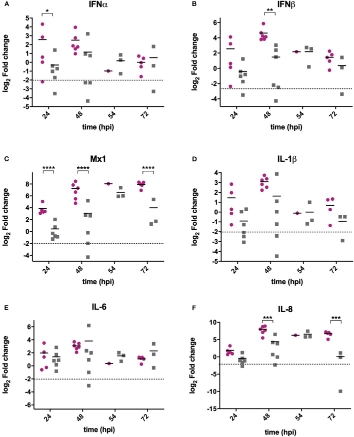
IBDV is economically important to the poultry industry. Very virulent (vv) strains cause higher mortality rates than other strains for reasons that remain poorly understood. In order to provide more information on IBDV disease outcome, groups of chickens (n = 18) were inoculated with the vv strain, UK661, or the classical strain, F52/70. Birds infected with UK661 had a lower survival rate (50%) compared to F52/70 (80%). There was no difference in peak viral replication in the bursa of Fabricius (BF), but the expression of chicken IFNα, IFNβ, MX1, and IL-8 was significantly lower in the BF of birds infected with UK661 compared to F52/70 (p < 0.05) as quantified by RTqPCR, and this trend was also observed in DT40 cells infected with UK661 or F52/70 (p < 0.05). The induction of expression of type I IFN in DF-1 cells stimulated with polyI:C (measured by an IFN-β luciferase reporter assay) was significantly reduced in cells expressing ectopic VP4 from UK661 (p < 0.05), but was higher in cells expressing ectopic VP4 from F52/70. Cells infected with a chimeric recombinant IBDV carrying the UK661-VP4 gene in the background of PBG98, an attenuated vaccine strain that induces high levels of innate responses (PBG98-VP4UK661) also showed a reduced level of IFNα and IL-8 compared to cells infected with a chimeric virus carrying the F52/70-VP4 gene (PBG98-VP4F52/70) (p < 0.01), and birds infected with PBG98-VP4UK661 also had a reduced expression of IFNα in the BF compared to birds infected with PBG98-VP4F52/70 (p < 0.05). Taken together, these data demonstrate that UK661 induced the expression of lower levels of anti-viral type I IFN and proinflammatory genes than the classical strain in vitro and in vivo and this was, in part, due to strain-dependent differences in the VP4 protein.

Keywords: IBDV; VP4; cytokines; inflammation; type I IFN; virulence.


Figures

Figure 1
Figure 1
The UK661 strain was more virulent than the F52/70 strain, but both strains replicated to the same peak titer in vivo. Birds were checked twice daily by two independent observers for clinical signs and a Kaplan Meier survival curve plotted of mock- (black), F52/70- (pink), and UK661- (gray) inoculated birds that reached their humane end points (clinical score of 11) (A). Clinical signs were quantified by a scoring system and divided into mild (1-7) and moderate (8-11). Each bird was assigned a clinical score at the indicated time points post-infection (B). Six birds per group were humanely culled at 24 and 48 h post-infection (hpi), one F52/70 and three UK661-infected birds reached their humane end-points at 54 hpi and the remaining birds were culled at 72 hpi. The bursa of Fabricius was harvested at necropsy and the log10 fold change in viral RNA copies/g tissue determined by RT-qPCR (C). The infectious titer was determined by titration onto DT40 cells in the method described by Reed and Muench. Virus titers were expressed as log10 TCID50/g of tissue (D). The horizontal lines are the mean values. Data passed a Shapiro-Wilk normality test before being analyzed by a one-way ANOVA and a Tukey's multiple comparison test (**p < 0.01).
Figure 2
Figure 2
The expression of type I IFN and pro-inflammatory genes was significantly reduced in BF tissue harvested from birds infected with strain UK661 compared to strain F52/70 in vivo. The bursa of Fabricius was harvested from mock and infected birds at necropsy and RNA extracted. RNA was reverse transcribed and amplified by quantitative PCR using specific primer sets for target genes. IFNα (A), IFNβ (B), Mx1 (C), IL-1β (D), IL-6 (E), and IL8 (F). The CT values were normalized to the housekeeping gene RPLPO and the log2 fold change in gene expression determined for the infected samples relative to the mock-infected samples in a ΔΔCT analysis and plotted for individual birds. The horizontal lines are the mean values. Data are representative of at least three replicate experiments and passed a Shapiro-Wilk normality test before being analyzed by a one-way ANOVA and a Tukey's multiple comparison test (*p < 0.05, **p < 0.01, ***p < 0.001, ****p < 0.0001). The dashed horizontal line represents the cut-off, below which genes were significantly down-regulated compared to mock-inoculated birds.
Figure 3
Figure 3
The expression of type I IFN and pro-inflammatory genes was significantly reduced in B cells infected with strain UK661 compared to strain F52/70 in vitro. DT40 Cells were infected at an MOI of 0.1 with either the UK661 or F52/70 IBDV strains, or mock-infected with media alone and RNA was extracted from the cells at the indicated time points post-infection. RNA was reverse transcribed and amplified by qPCR using specific primer sets. The CT values were normalized to the housekeeping gene RPLPO and the log10 fold change in virus gene expression determined for the infected samples relative to the mock-infected samples in a ΔΔCT analysis and plotted (A). The log2 fold change in host-cell gene expression was also determined for the infected samples relative to the mock-infected samples in a ΔΔCT analysis and plotted (B–G). Data subsequently passed a Shapiro-Wilk normality test before being analyzed by a one-way ANOVA and a Tukey's multiple comparison test (*P < 0.05) (A), or a two-tailed unpaired Student's t-test (*p < 0.05, ***p < 0.001). Data shown are representative of at least three replicate experiments, columns represent the mean values, and error bars represent the standard deviation of the mean.
Figure 4
Figure 4
The VP4 protein from the UK661 strain antagonized IFNβ induction, but the VP4 protein from the F52/70 strain did not in vitro. DF-1s were transfected with the chicken IFNβ promoter Firefly luciferase reporter and a constitutively active Renilla expression plasmid and 500 ng of either eGFP-UK661-VP4 or eGFP-F52/70-VP4 expression plasmids, or a control plasmid expressing eGFP alone. Twenty-four hours post-transfection, cells were re-transfected with poly I:C. At 6 h post-transfection, cells were lysed and luciferase activity quantified. Firefly luciferase activity was normalized to Renilla expression. Data presented are the means of three independent experiments and passed a Shapiro-Wilk normality test before being analyzed by a one-way ANOVA and a Tukey's multiple comparison test (*p < 0.05, **p < 0.01). Error bars represent the standard error of the mean (SEM). In a parallel experiment, transfected cells were lysed and samples denatured and subject to SDS-PAGE gel electrophoresis followed by transfer to a nitrocellulose membrane and staining with anti-VP4 and anti-β-actin antibodies in a western blot.
Figure 5
Figure 5
The PBG98-VP4 UK661 virus inhibits the induction of IFNα and IL-8 to a greater extent than the PBG98-VP4F52/70 virus in vitro. DF-1 cells were infected with PBG98, PBG98-VP4UK661, and PBG98-VP4F52/70 viruses at an MOI of 1, before RNA was extracted at the indicated time points post-infection and reverse transcribed. Virus specific primers were used to amplify the cDNA by quantitative (q)PCR, the CT values were normalized to the housekeeping gene RPLPO and the log10 fold change in virus gene expression was determined for the infected samples relative to the mock-infected controls in a ΔΔCT analysis and plotted. A one-way ANOVA and a Tukey's multiple comparison test was performed where no significant difference was found at any time point between the three viruses (A). A panel of genes, IFNα (B), IFNβ (C), Mx1 (D), IL-1β (E), and IL-8 (F), were amplified by qPCR using specific primer sets for target genes, before the CT values were normalized to the housekeeping gene RPLPO and the log2 fold change in gene expression determined for the infected samples relative to the mock-infected controls in a ΔΔCT analysis and plotted. Data are representative of at least three replicate experiments and passed a Shapiro-Wilk normality test before being analyzed by a one-way ANOVA and a Tukey's multiple comparison test (*p < 0.05, **p < 0.01, ***p < 0.001, ****p < 0.0001). The mean values are plotted and the error bars are the standard error of the mean (SEM). The dashed horizontal line represents the cut-off, below which genes were significantly down-regulated.
Figure 6
Figure 6
The PBG98-VP4 UK661 virus inhibits the induction of IFNα to a greater extent than the PBG98-VP4F52/70 virus in vivo. Birds were inoculated with 1.8 × 10³ TCID50 of the PBG98, PBG98-VP4UK661, and PBG98-VP4F52/70 viruses, and the bursa of Fabricius was harvested at necropsy from 6 birds per group at 2, 4, and 14 days post-inoculation. RNA was extracted prior to reverse transcription to cDNA and qPCR amplification with virus-specific primers. CT values were normalized to a housekeeping gene and expressed as log10fold change viral RNA relative to mock-infected samples as per the ΔΔCT method. The data passed a Shapiro-Wilk normality test before being analyzed by a one-way ANOVA and a Tukey's multiple comparison test (not significant) (A). At 2 and 4 days post-inoculation, cDNA was amplified by qPCR for a panel genes: IFNα (B), IFNβ (C), Mx1 (D), IL-1β (E), and IL-8 (F). The CT values were normalized to the housekeeping gene RPLPO and expressed relative to mock-infected samples using the ΔΔCT method. Data are representative of at least three replicate experiments and passed a Shapiro-Wilk normality test before being analyzed by a one-way ANOVA and a Tukey's multiple comparison test (*p < 0.05, ***p < 0.001). Horizontal lines represent the mean and error bars represent the standard error of the mean (SEM).

References

    1. Baba T. W., Giroir B. P., Humphries E. H. (1985). Cell lines derived from avian lymphomas exhibit two distinct phenotypes. Virology 144, 139–151. 10.1016/0042-6822(85)90312-5 - DOI - PubMed
    1. Bayliss C. D., Spies U., Shaw K., Peters R. W., Papageorgiou A., Muller H., et al. . (1990). A comparison of the sequences of segment A of four infectious bursal disease virus strains and identification of a variable region in VP2. J. Gen. Virol. 71 (Pt 6), 1303–1312. 10.1099/0022-1317-71-6-1303 - DOI - PubMed
    1. Brown M. D., Green P., Skinner M. A. (1994). VP2 sequences of recent European ‘very virulent’ isolates of infectious bursal disease virus are closely related to each other but are distinct from those of ‘classical’ strains. J. Gen. Virol. 75 (Pt 3), 675–680. 10.1099/0022-1317-75-3-675 - DOI - PubMed
    1. Brown M. D., Skinner M. A. (1996). Coding sequences of both genome segments of a European ‘very virulent’ infectious bursal disease virus. Virus Res. 40, 1–15. 10.1016/0168-1702(95)01253-2 - DOI - PubMed
    1. Campbell E. A., Reddy V. R. A. P., Gray A. G., Wells J., Simpson J., Skinner M. A., et al. . (2020). Discrete virus factories form in the cytoplasm of cells co-infected with two replication competent tagged reporter birnaviruses, that subsequently coalesce over time. J. Virol. 10.1128/JVI.02107-19. [Epub ahead of print]. - DOI - PMC - PubMed

Publication types

Substances