Review
. 2020 Jun 3:8:409.
doi: 10.3389/fcell.2020.00409. eCollection 2020.

MicroRNAs: From Mechanism to Organism

Affiliations
Review

MicroRNAs: From Mechanism to Organism

Philipp J Dexheimer et al. Front Cell Dev Biol. .

Abstract

MicroRNAs (miRNAs) are short, regulatory RNAs that act as post-transcriptional repressors of gene expression in diverse biological contexts. The emergence of small RNA-mediated gene silencing preceded the onset of multicellularity and was followed by a drastic expansion of the miRNA repertoire in conjunction with the evolution of complexity in the plant and animal kingdoms. Along this process, miRNAs became an essential feature of animal development, as no higher metazoan lineage tolerated loss of miRNAs or their associated protein machinery. In fact, ablation of the miRNA biogenesis machinery or the effector silencing factors results in severe embryogenesis defects in every animal studied. In this review, we summarize recent mechanistic insight into miRNA biogenesis and function, while emphasizing features that have enabled multicellular organisms to harness the potential of this broad class of repressors. We first discuss how different mechanisms of regulation of miRNA biogenesis are used, not only to generate spatio-temporal specificity of miRNA production within an animal, but also to achieve the necessary levels and dynamics of expression. We then explore how evolution of the mechanism for small RNA-mediated repression resulted in a diversity of silencing complexes that cause different molecular effects on their targets. Multicellular organisms have taken advantage of this variability in the outcome of miRNA-mediated repression, with differential use in particular cell types or even distinct subcellular compartments. Finally, we present an overview of how the animal miRNA repertoire has evolved and diversified, emphasizing the emergence of miRNA families and the biological implications of miRNA sequence diversification. Overall, focusing on selected animal models and through the lens of evolution, we highlight canonical mechanisms in miRNA biology and their variations, providing updated insight that will ultimately help us understand the contribution of miRNAs to the development and physiology of multicellular organisms.

Keywords: Argonaute; Dicer; Drosha; biogenesis; development; evolution; miRNA; silencing.


Figures

FIGURE 1
FIGURE 1
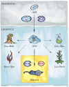
The evolutionary origins of miRNAs in eukaryotes. Two key players in small RNA-mediated silencing, Argonaute proteins and RNase III like enzymes, originated in prokaryotes. The miRNA pathway in animals emerged with the birth of the Microprocessor, composed of Drosha and Pasha, in unicellular holozoans (Bråte et al., 2018). Diverse lineages that branched from the last eukaryotic common ancestor (LECA) also evolved miRNA-like pathways. However, it is still under debate whether these evolved independently in four additional clades: Slime molds, Green algae, Brown Algae, and Land plants (Kruse et al., 2016; Valli et al., 2016; Cock et al., 2017; Bråte et al., 2018); or if the pathway was already present in the last common ancestor (Moran et al., 2017).
FIGURE 2
FIGURE 2
Schematic of the miRNA biogenesis pathway. The different steps leading to production of a mature miRNA are shown. Kinetic studies in Drosophila revealed that biogenesis is fast for most miRNAs but loading into Argonaute represents the rate-limiting step (based on Reichholf et al., 2019).
FIGURE 3
FIGURE 3
Canonical miRNA-silencing mechanism in animals. miRNAs elicit repression of target genes usually through a combination of translational repression and promotion of mRNA decay. Argonaute is guided by a miRNA to a cognate target mRNA and tethers GW182, forming the core of the most common animal miRISC. GW182 interacts with PABP and recruits the deadenylase complexes CCR4-NOT or Pan2-Pan3 (not shown), leading to deadenylation, decapping and ultimately exonucleolytic decay. Inhibition of translation occurs mainly at the initiation step by interfering with assembly or activity of eIF4F, via eIF4E-T, and DDX6.
FIGURE 4
FIGURE 4
Alternative outcomes of miRNA-mediated targeting. In addition to the common miRISC effects on translation and stability of target mRNAs, other functional outcomes of miRISC binding to an mRNA are possible. (A) Full sequence complementarity results in Ago-mediated target cleavage, a mechanism that resembles the mode of action commonly employed in cnidaria and plants. (B) Target-mediated miRNA degradation is induced by interaction with targets through extensive pairing, in particular extending to the miRNA 3′ end. (C) Recruitment of Argonaute in the absence of GW182 results in inhibition of translation without affecting mRNA stability (likely involving alternative co-factors).
FIGURE 5
FIGURE 5
Diversification of the metazoan miRNA repertoire. Shown are numbers of individual miRNAs/miRNA-families (in bold) annotated with high confidence for various clades (https://mirgenedb.org, Fromm et al., 2020). Number of miRNA families present in the last common ancestors of branching clades are noted above the split. Exemplary organisms depicted are Amphimedon queenslandica, Nematostella vectensis, Caenorhabditis elegans, Drosophila melanogaster, Danio rerio, Mus musculus, and Homo sapiens. Data for bilateria is derived from Fromm et al. (2020), for Amphimedon and Nematostella numbers were retrieved from the work of Grimson et al. (2008); Moran et al. (2014), and Calcino et al. (2018).

References

    1. Abbott A. L., Alvarez-Saavedra E., Miska E. A., Lau N. C., Bartel D. P., Horvitz H. R., et al. (2005). The let-7 MicroRNA family members mir-48, mir-84, and mir-241 function together to regulate developmental timing in Caenorhabditis elegans. Dev. Cell 9 403–414. 10.1016/j.devcel.2005.07.009 - DOI - PMC - PubMed
    1. Agarwal V., Subtelny A. O., Thiru P., Ulitsky I., Bartel D. P. (2018). Predicting microRNA targeting efficacy in Drosophila. Genome Biol. 19:152. 10.1186/s13059-018-1504-3 - DOI - PMC - PubMed
    1. Alberti C., Cochella L. (2017). A framework for understanding the roles of miRNAs in animal development. Development 144 2548–2559. 10.1242/dev.146613 - DOI - PubMed
    1. Alberti C., Manzenreither R. A., Sowemimo I., Burkard T. R., Wang J., Mahofsky K., et al. (2018). Cell-type specific sequencing of microRNAs from complex animal tissues. Nat. Methods 15 283–289. 10.1038/nmeth.4610 - DOI - PMC - PubMed
    1. Alisch R. S., Jin P., Epstein M., Caspary T., Warren S. T. (2007). Argonaute2 is essential for mammalian gastrulation and proper mesoderm formation. PLoS Genet. 3:e227. 10.1371/journal.pgen.0030227 - DOI - PMC - PubMed

LinkOut - more resources