

Induction of Autophagy by Pterostilbene Contributes to the Prevention of Renal Fibrosis via Attenuating NLRP3 Inflammasome Activation and Epithelial-Mesenchymal Transition
- PMID: 32582712
- PMCID: PMC7283393
- DOI: 10.3389/fcell.2020.00436
Induction of Autophagy by Pterostilbene Contributes to the Prevention of Renal Fibrosis via Attenuating NLRP3 Inflammasome Activation and Epithelial-Mesenchymal Transition
Abstract
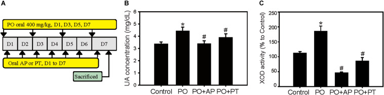
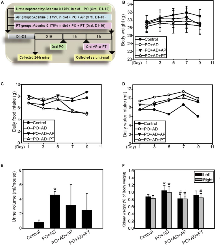
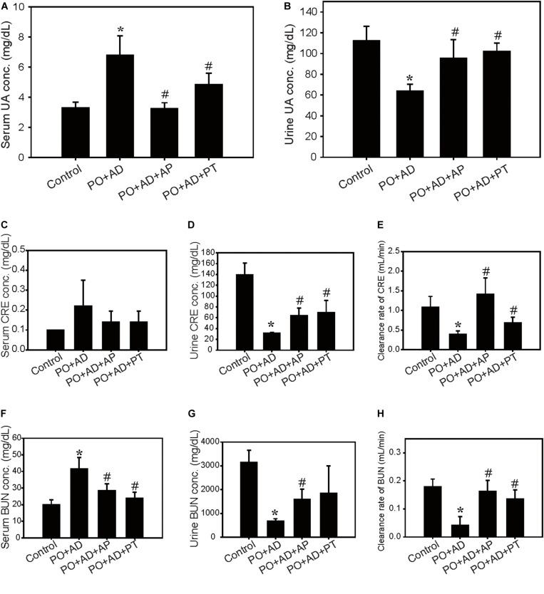
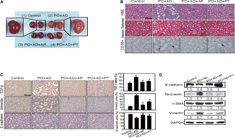
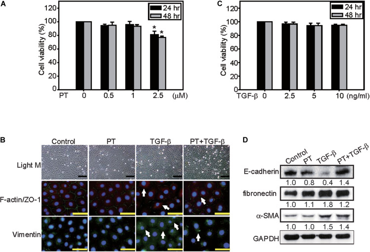
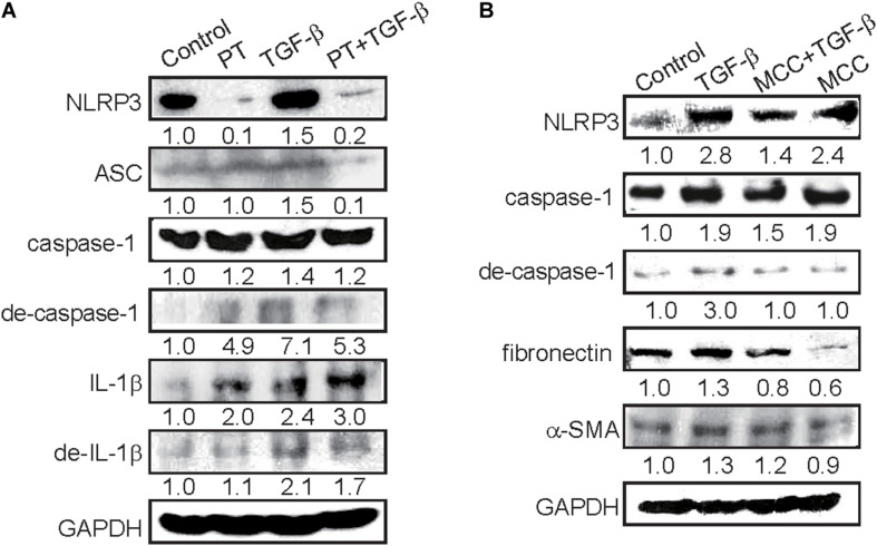
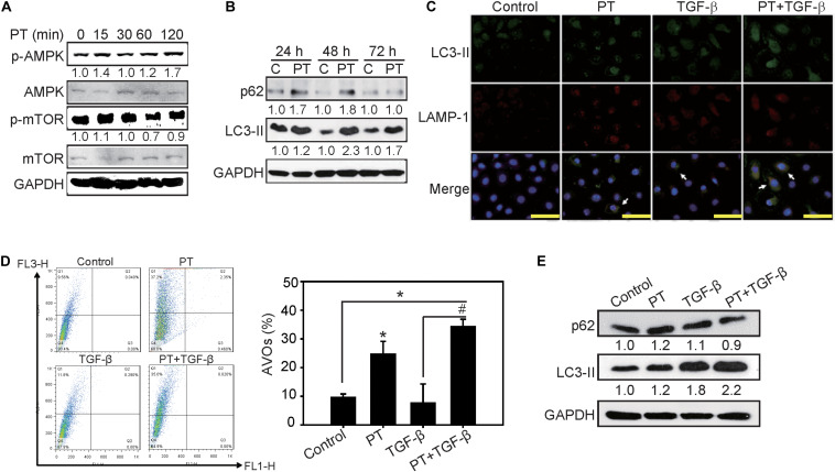
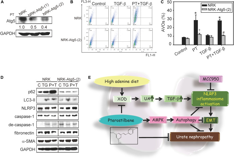
Chronic kidney disease (CKD) is recognized as a global public health problem. NLRP3 inflammasome activation has been characterized to mediate diverse aspect mechanisms of CKD through regulation of proinflammatory cytokines, tubulointerstitial injury, glomerular diseases, renal inflammation, and fibrosis pathways. Autophagy is a characterized negative regulation mechanism in the regulation of the NLRP3 inflammasome, which is now recognized as the key regulator in the pathogenesis of inflammation and fibrosis in CKD. Thus, autophagy is undoubtedly an attractive target for developing new renal protective treatments of kidney disease via its potential effects in regulation of inflammasome. However, there is no clinical useful agent targeting the autophagy pathway for patients with renal diseases. Pterostilbene (PT, trans-3,5-dimethoxy-4-hydroxystilbene) is a natural analog of resveratrol that has various health benefits including autophagy inducing effects. Accordingly, we aim to investigate underlying mechanisms of preventive and therapeutic effects of PT by reducing NLRP3 inflammasome activation and fibrosis through autophagy-inducing effects. The renal protective effects of PT were evaluated by potassium oxonate (PO)-induced hyperuricemia and high adenine diet-induced CKD models. The autophagy induction mechanisms and anti-fibrosis effects of PT by down-regulation of NLRP3 inflammasome are investigated by using immortalized rat kidney proximal tubular epithelial NRK-52E cells. To determine the role of autophagy induction in the alleviating of NLRP3 inflammasome activation and epithelial-mesenchymal transition (EMT), NRK-52E with Atg5 knockdown [NRK-Atg5-(2)] cells were applied in the study. The results indicated that PT significantly reduces serum uric acid levels, liver xanthine oxidase activity, collagen accumulation, macrophage recruitment, and renal fibrosis in CKD models. At the molecular levels, pretreatment with PT downregulating TGF-β-triggered NLRP3 inflammasome activation, and subsequent EMT in NRK-52E cells. After blockage of autophagy by treatment of Atg5 shRNA, PT loss of its ability to prevent NLRP3 inflammasome activation and EMT. Taken together, we suggested the renal protective effects of PT in urate nephropathy and proved that PT induces autophagy leading to restraining TGF-β-mediated NLRP3 inflammasome activation and EMT. This study is also the first one to provide a clinical potential application of PT for a better management of CKD through its autophagy inducing effects.
Keywords: NLRP3 inflammasome; autophagy; epithelial-mesenchymal transition; pterostilbene; renal fibrosis.
Copyright © 2020 Wang, Chen, Hsiao, Pan, Wang, Chen, Ho, Huang and Chen.
Figures
Similar articles
-
Complanatoside A targeting NOX4 blocks renal fibrosis in diabetic mice by suppressing NLRP3 inflammasome activation and autophagy.Phytomedicine. 2022 Sep;104:154310. doi: 10.1016/j.phymed.2022.154310. Epub 2022 Jul 5. Phytomedicine. 2022. PMID: 35843189
-
Contribution of TGF-Beta-Mediated NLRP3-HMGB1 Activation to Tubulointerstitial Fibrosis in Rat With Angiotensin II-Induced Chronic Kidney Disease.Front Cell Dev Biol. 2020 Feb 5;8:1. doi: 10.3389/fcell.2020.00001. eCollection 2020. Front Cell Dev Biol. 2020. PMID: 32117956 Free PMC article.
-
Weicao capsule ameliorates renal injury through increasing autophagy and NLRP3 degradation in UAN rats.Int J Biochem Cell Biol. 2018 Mar;96:1-8. doi: 10.1016/j.biocel.2018.01.001. Epub 2018 Jan 2. Int J Biochem Cell Biol. 2018. PMID: 29305119
-
The Role of NLRP3 Inflammasome Activation in the Epithelial to Mesenchymal Transition Process During the Fibrosis.Front Immunol. 2020 May 20;11:883. doi: 10.3389/fimmu.2020.00883. eCollection 2020. Front Immunol. 2020. PMID: 32508821 Free PMC article. Review.
-
Mechanistic insights into hyperuricemia-associated renal abnormalities with special emphasis on epithelial-to-mesenchymal transition: Pathologic implications and putative pharmacologic targets.Pharmacol Res. 2020 Nov;161:105209. doi: 10.1016/j.phrs.2020.105209. Epub 2020 Sep 23. Pharmacol Res. 2020. PMID: 32979505 Review.
Cited by
-
Alleviation of Hyperuricemia by Strictinin in AML12 Mouse Hepatocytes Treated with Xanthine and in Mice Treated with Potassium Oxonate.Biology (Basel). 2023 Feb 17;12(2):329. doi: 10.3390/biology12020329. Biology (Basel). 2023. PMID: 36829604 Free PMC article.
-
Skin damage induced by zinc oxide nanoparticles combined with UVB is mediated by activating cell pyroptosis via the NLRP3 inflammasome-autophagy-exosomal pathway.Part Fibre Toxicol. 2022 Jan 5;19(1):2. doi: 10.1186/s12989-021-00443-w. Part Fibre Toxicol. 2022. PMID: 34983566 Free PMC article.
-
Pterostilbene Protects against Osteoarthritis through NLRP3 Inflammasome Inactivation and Improves Gut Microbiota as Evidenced by In Vivo and In Vitro Studies.J Agric Food Chem. 2024 Apr 16;72(16):9150-63. doi: 10.1021/acs.jafc.3c09749. Online ahead of print. J Agric Food Chem. 2024. PMID: 38624135 Free PMC article.
-
LncRNA TUG1 promotes pulmonary fibrosis progression via up-regulating CDC27 and activating PI3K/Akt/mTOR pathway.Epigenetics. 2023 Dec;18(1):2195305. doi: 10.1080/15592294.2023.2195305. Epigenetics. 2023. PMID: 36994860 Free PMC article.
-
Prospective Pharmacological Potential of Resveratrol in Delaying Kidney Aging.Int J Mol Sci. 2021 Jul 31;22(15):8258. doi: 10.3390/ijms22158258. Int J Mol Sci. 2021. PMID: 34361023 Free PMC article. Review.
References
-
- Chen R. J., Chen M. H., Chen Y. L., Hsiao C. M., Chen H. M., Chen S. J., et al. (2017a). Evaluating the urate-lowering effects of different microbial fermented extracts in hyperuricemic models accompanied with a safety study. J. Food Drug Anal. 25 597–606. 10.1016/j.jfda.2016.07.003 - DOI - PMC - PubMed
-
- Chen R. J., Wu P. H., Ho C. T., Way T. D., Pan M. H., Chen H. M., et al. (2017c). P53-dependent downregulation of hTERT protein expression and telomerase activity induces senescence in lung cancer cells as a result of pterostilbene treatment. Cell Death Dis. 8:e2985. 10.1038/cddis.2017.333 - DOI - PMC - PubMed
Grants and funding
LinkOut - more resources
Full Text Sources
Other Literature Sources