. 2020 Jan;8(4):419-423.

C-MAF Expression in Adult Human Ocular Surface and its Implication in Pterygium Pathogenesis

Affiliations

C-MAF Expression in Adult Human Ocular Surface and its Implication in Pterygium Pathogenesis

Hasan Mahmud Reza et al. Rep Biochem Mol Biol. 2020 Jan.

Abstract

Background: c-MAF, a transcription factor that belongs to the b-Zip Maf transcription factor family, was found to be critical for lens development in vertebrates. It is a well-known fact that the adult human ocular surface expresses c-MAF, however, its role in the limbus, cornea and conjunctiva remains unknown. Thus, the present study aimed to investigate c-MAF expression within the human ocular surface, and its potential role in pterygium pathogenesis.

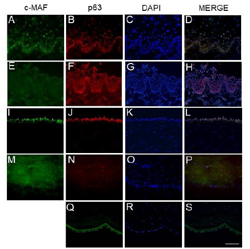
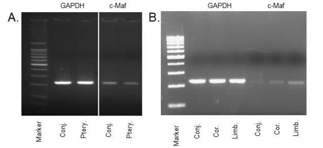
Methods: We performed immunohistochemical staining to detect c-MAF expression in frozen adult human tissue sections, including the limbus, cornea and conjunctiva, and cultured cells from eye cadavers. We then compared c-MAF expression to the expression of a known protein, P63. Lastly, we performed RT-PCR, and immunohistochemistry for c-MAF expression in healthy adult human conjunctiva and pterygium.

Results: We found differential c-MAF expression between adult human limbus, cornea and conjunctiva tissues. Further, we observed that c-MAF is downregulated in the pterygium compared to healthy conjunctiva.

Conclusion: Overall, our results suggest that c-MAF may play a context-specific role in maintaining limbal, corneal and conjunctival homeostasis, and may be critical for preventing pterygium development in humans.

Keywords: C-MAF Expression; Conjunctiva; Human Ocular Surface; Pterygium.


Figures

Fig. 1
Fig. 1
Immunofluorescent staining of c-MAF and p63 on frozen sections of human limbal epithelial crypt (A-D), palisades of Vogt of limbus (E-H), peripheral cornea (I-L), corneal stroma (M-P) and endothelial cells (Q-S). DAPI stains cell nuclei (3rd column). Limbal epithelial crypt shows equal expression of c-MAF (A, D) and p63 (B, D). Cells in the palisades of Vogt express p63 (F, H) but not c-MAF (E, H). Only the basal layers of peripheral cornea show c-MAF (I, L) and p63 (J, L) expression. Some stromal cells are also positive to c-MAF (M, P) and p63 (N, P) reactivity. Endothelial cells clearly exhibit c-MAF expression (Q, S).
Fig. 2
Fig. 2
Gene expression profiling in cultured human limbal epithelial cells. Strong expression of c-MAF (A-C) and p63 (E-G) is found in central cells of the colonies. Ki67 is detected in almost all cells (I-K). CK15 is strongly expressed in peripheral cells (H). E-Cadherin expression is strong in central cells of the colonies (L). Phase contrast image shows cell morphology of the clone (D). DAPI depicts cell nuclei (B, F, & G).
Fig. 3
Fig. 3
Expression pattern of c-MAF and p63 in human conjunctival tissue (A, D) and cultured cells (E-H). c-MAF is expressed in some cells in the most upper layer (A, D, arrowheads) while weakly expressed in a subset of population with small nuclei in the basal layer (A, D, arrows). p63 in strongly expressed in basal layer (B, D). Although c-MAF is expressed in cultured conjunctival cells (E, H), p63 is absent (F). DAPI depicts cell nuclei (C, G).
Fig. 4
Fig. 4
Expression of c-MAF and Ki67 in cultured conjunctival as well as pterygial cells. c-MAF expression is stronger in normal conjunctival cells (A-C) than pteyrigium cells (D-F). Differentiation marker Ki67 is strongly expressed in most cells of the pterygium (J-L) but weakly expressed in conjunctiva (G-I). Cell morphology appears same in both cases (M, N). DAPI stains cell nuclei (2nd column).
Fig. 5
Fig. 5
RT-PCR shows the differential expression of c-MAF in conjunctival and pterygial tissues (A) and in normal ocular surface (B, limbus, cornea, conjunctiva).

Similar articles

Cited by

References

    1. Swamynathan SK. Ocular surface development and gene expression. J ophthalmol. 2013;2013:103947. - PMC - PubMed
    1. Schermer A, Galvin S, Sun TT. Differentiation-related expression of a major 64K corneal keratin in vivo and in culture suggests limbal location of corneal epithelial stem cells. J Cell Biol . 1986;103(1):49–62. - PMC - PubMed
    1. Kim JI, Li T, Ho IC, Grusby MJ, Glimcher LH. Requirement for the c-MAF transcription factor in crystallin gene regulation and lens development. . Proc Natl Acad Sci U S A . 1999;96(7):3781–3785. - PMC - PubMed
    1. Kienast J, Berdel WE. c-MAF in multiple myeloma: an oncogene enhancing tumor-stroma interactions. Cancer cell . 2004;5(2):109–110. - PubMed
    1. Chui J, Coroneo MT, Tat LT, Crouch R, Wakefield D, Di Girolamo N. Ophthalmic pterygium: a stem cell disorder with premalignant features. . Am J Pathol . 2011;178(2):817–827. - PMC - PubMed

LinkOut - more resources