. 2020 Jun 12;6(24):eaba1430.
doi: 10.1126/sciadv.aba1430. eCollection 2020 Jun.

GABAergic interneurons excite neonatal hippocampus in vivo

Affiliations

GABAergic interneurons excite neonatal hippocampus in vivo

Yasunobu Murata et al. Sci Adv. .

Abstract

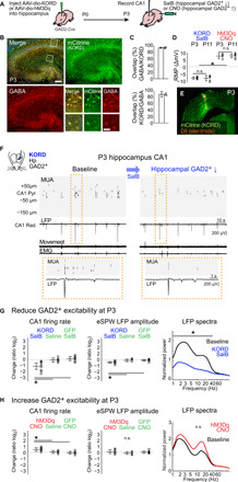
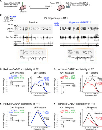
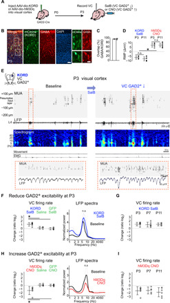
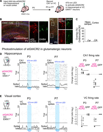
GABAergic interneurons are proposed to be critical for early activity and synapse formation by directly exciting, rather than inhibiting, neurons in developing hippocampus and neocortex. However, the role of GABAergic neurons in the generation of neonatal network activity has not been tested in vivo, and recent studies have challenged the excitatory nature of early GABA. By locally manipulating interneuron activity in unanesthetized neonatal mice, we show that GABAergic neurons are excitatory in CA1 hippocampus at postnatal day 3 (P3) and are responsible for most of the spontaneous firing of pyramidal cells at that age. Hippocampal interneurons become inhibitory by P7, whereas visual cortex interneurons are already inhibitory by P3 and remain so throughout development. These regional and age-specific differences are the result of a change in chloride reversal potential, because direct activation of light-gated anion channels in glutamatergic neurons drives CA1 firing at P3, but silences it at P7 in CA1, and at all ages in visual cortex. This study in the intact brain reveals that GABAergic interneuron excitation is essential for network activity in neonatal hippocampus and confirms that visual cortical interneurons are inhibitory throughout early postnatal development.


Figures

Fig. 1
Fig. 1. GABAergic interneurons are excitatory in 3-day-old hippocampus in vivo.
(A) Experimental design. (B) Colabeling of AAV-dF-KORD-IRES-mCitrine expressed in GAD2-Cre mouse with anti-GABA. Scale bars, 100 and 20 μm. (C) Percentage of mCitrine(KORD)–expressing neurons coexpressing GABA and percentage of GABA-expressing neurons coexpressing mCitrine(KORD) (mean ± 95% CI, n = 3, 3). (D) Change in membrane potential in hippocampal slices at P3 and P11. SalB hyperpolarized KORD–expressing neurons and CNO depolarized hM3Dq–expressing neurons at both ages (n = 6, 6 , 5, and 7; ANOVA, P < 0.001). (E) Representative localization of electrode and viral expression in P3 animal. (F) Representative recording for P3 reduction of GABAergic neuron excitability. MUA of spontaneous activity in CA1 hippocampus, along with associated stratum radiatum LFP and thoracic movement detection and electromyography. Activity is dominated by early sharp waves (eSPW) whose spike density is reduced following subcutaneous SalB (KORD agonist) injection. (G) Quantification of KORD-induced suppression of GABAergic neuron excitability and control conditions. [Pyramidal cell layer firing rate (n = 10, 6, and 8; ANOVA, P < 0.001), eSPW LFP amplitude (n = 10, 6, and 8; P = 0.002), and normalized (to mean of 1- to 100-Hz baseline) spectral power for stratum radiatum LFP, n = 10]. (H) Quantification of hM3Dq-induced increase in GABAergic excitability (n = 7, 6, and 6; P = 0.005; P = 0.33; n = 7). All values and statistics are listed in table S1.
Fig. 2
Fig. 2. Hippocampal GABAergic neurons are inhibitory by P7.
(A) Experimental design. (B) Representative recording for GABAergic neuron suppression in P7 hippocampus. (C and D) Quantification of suppression (C) and enhancement (D) of GABAergic neuron excitability at P7 [(C): CA1 firing rate: KORD-SalB: 1.14 ± 0.62 (n = 5), KORD-saline: 0.04 ± 0.35 (n = 4), GFP-SalB: −0.04 ± 0.43 (n = 4), P = 0.001; LFP spectra: P < 0.05 at 6.9 to 14.7 Hz (n = 5); (D): CA1 firing rate: hM3Dq-CNO: −2.37 ± 2.02 (n = 5), hM3Dq-saline: 0.17 ± 0.21 (n = 4), GFP-CNO: 0.13 ± 0.43 (n = 5), P = 0.003; LFP spectra: P < 0.05 at 7.7 to 93.3 Hz (n = 5)]. (E and F) Similar quantification at P11 [(E): CA1 firing rate: KORD-SalB: 1.11 ± 0.57 (n = 6), KORD-saline: 0.12 ± 0.22 (n = 8), GFP-SalB: −0.03 ± 0.28 (n = 4), P < 0.001; LFP spectra: not significant (n.s.) (n = 6); (F): CA1 firing rate, hM3Dq-CNO: −2.09 ± 1.29 (n = 7), hM3Dq-saline: 0.18 ± 0.22 (n = 5), GFP-CNO: −0.07 ± 0.37 (n = 5), P = 0.001; LFP spectra, P < 0.05 at 2.3 to 93.3 Hz (n = 7)].
Fig. 3
Fig. 3. GABAergic neurons in visual cortex have a net inhibitory action as early as P3.
(A) Experimental design. (B and C) Colocalization of KORD expression with GABA [93.7 ± 6.7% (n = 3)]. Scale bars, 50 and 10 μm. (D) Change in membrane potential of GAD2+ neurons by activation of KORD and hM3Dq at P3 and P11 in visual cortical slices [P3 KORD: −6.62 ± 1.88 (n = 5), P11 KORD: −8.03 ± 2.14 (n = 7), P3 hM3Dq: 7.18 ± 2.23 (n = 6), P11 hM3Dq: 7.23 ± 2.06 (n = 8), P < 0.001]. (E) Representative recording of visual cortex at P3 and the effect of GABAergic neuron suppression. LFP spectrogram is from the presumptive input layer. (F) Quantification of change in superficial layer firing rate [KORD-SalB: 1.25 ± 0.5 (n = 6), KORD-saline: 0.22 ± 0.4 (n = 4), GFP-SalB: −0.03 ± 0.35 (n = 5), P = 0.001] and LFP spectral power [n.s. (n = 6)] following suppression of GABAergic neuron excitability by KORD activation. (G) Firing rate change at P3, P7, and P11 to KORD activation [P3: 1.25 ± 0.5 (n = 6), P7: 1.11 ± 0.71 (n = 6), P11: 1.17 ± 0.43 (n = 5); P = 0.89]. (H and I) As (F) and (G) but for GABAergic neuron enhancement by hM3Dq activation [(H): VC firing rate: hM3Dq-CNO: −1.51 ± 1.18 (n = 6), hM3Dq-saline: 0.11 ± 0.46 (n = 4), GFP-CNO: 0.02 ± 0.32 (n = 5), P = 0.007; LFP spectra: n.s. (n = 6); (I): P3: −1.51 ± 1.18 (n = 6), P7: −1.43 ± 1.02 (n = 5), P11: −1.94 ± 1.35 (n = 6), P = 0.71].
Fig. 4
Fig. 4. Changing role of anion conductance likely underlies the regional and age heterogeneity of GABA function.
(A to C) stGtACR2, an anion-conducting channelrhodopsin with a soma-targeting motif, is virally expressed in non-GABAergic neurons in hippocampus or cortex of EMX1-Cre mice [(C): hippocampus: 8.5 ± 8.5% (n = 3); visual cortex: 10.7 ± 7.6% (n = 3)]. Scale bars, 50 and 10 μm. (D) Photostimulation of stGtACR2 in hippocampal glutamatergic neurons (470 nm LED, 1 s) increased CA1 firing at P3 but decreases it at P7 [P3 stGtACR2: 0.74 ± 0.38 (n = 5), P3 GFP: −0.07 ± −0.35 (n = 5), P = 0.001; P7 stGtACR2: −3.51 ± −4.25 (n = 7), P7 GFP: 0.04 ± −0.01 (n = 5), P = 0.001]. (E) In visual cortex, photostimulation of stGtACR2 in glutamatergic neurons decreased MUA at both P3 and P7 [P3 stGtACR2: −2.65 ± −4.68 (n = 4), P3 GFP: 0.08 ± −0.29 (n = 5), P = 0.002; P7 stGtACR2: −2.43 ± −3.27 (n = 4), P7 GFP: 0.18 ± 0.04 (n = 3), P = 0.001].

Similar articles

Cited by

References

    1. Che A., Babij R., Iannone A. F., Fetcho R. N., Ferrer M., Liston C., Fishell G., De Marco García N. V., Layer I interneurons sharpen sensory maps during neonatal development. Neuron 99, 98–116.e7 (2018). - PMC - PubMed
    1. Le Magueresse C., Monyer H., GABAergic interneurons shape the functional maturation of the cortex. Neuron 77, 388–405 (2013). - PubMed
    1. Tuncdemir S. N., Wamsley B., Stam F. J., Osakada F., Goulding M., Callaway E. M., Rudy B., Fishell G., Early somatostatin interneuron connectivity mediates the maturation of deep layer cortical circuits. Neuron 89, 521–535 (2016). - PMC - PubMed
    1. Duan Z. R. S., Che A., Chu P., Modol L., Bollmann Y., Babij R., Fetcho R. N., Otsuka T., Fuccillo M. V., Liston C., Pisapia D. J., Cossart R., De Marco García N. V., GABAergic restriction of network dynamics regulates interneuron survival in the developing cortex. Neuron 105, 75–92.e5 (2020). - PMC - PubMed
    1. Marques-Smith A., Lyngholm D., Kaufmann A.-K., Stacey J. A., Hoerder-Suabedissen A., Becker E. B. E., Wilson M. C., Molnár Z., Butt S. J. B., A transient translaminar gabaergic interneuron circuit connects thalamocortical recipient layers in neonatal somatosensory cortex. Neuron 89, 536–549 (2016). - PMC - PubMed

Publication types