. 2020 Nov 5;136(19):2151-2161.
doi: 10.1182/blood.2020005827.

Lysine-specific demethylase 1A restricts ex vivo propagation of human HSCs and is a target of UM171

Affiliations

Lysine-specific demethylase 1A restricts ex vivo propagation of human HSCs and is a target of UM171

Agatheeswaran Subramaniam et al. Blood. .

Abstract

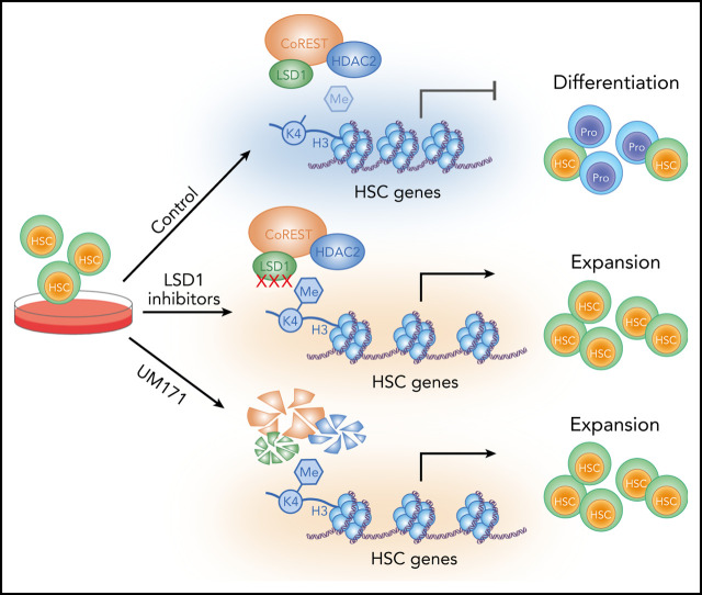
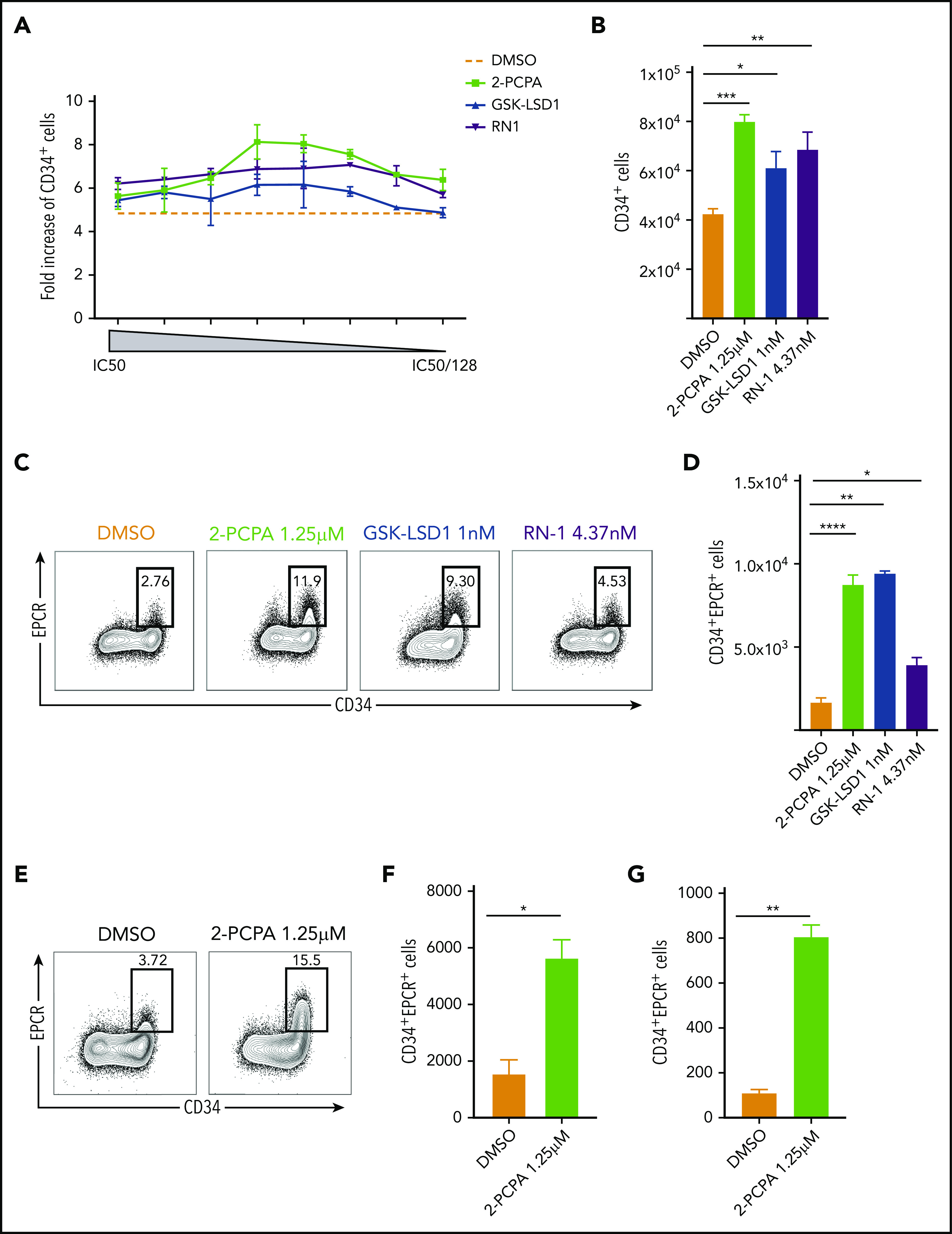
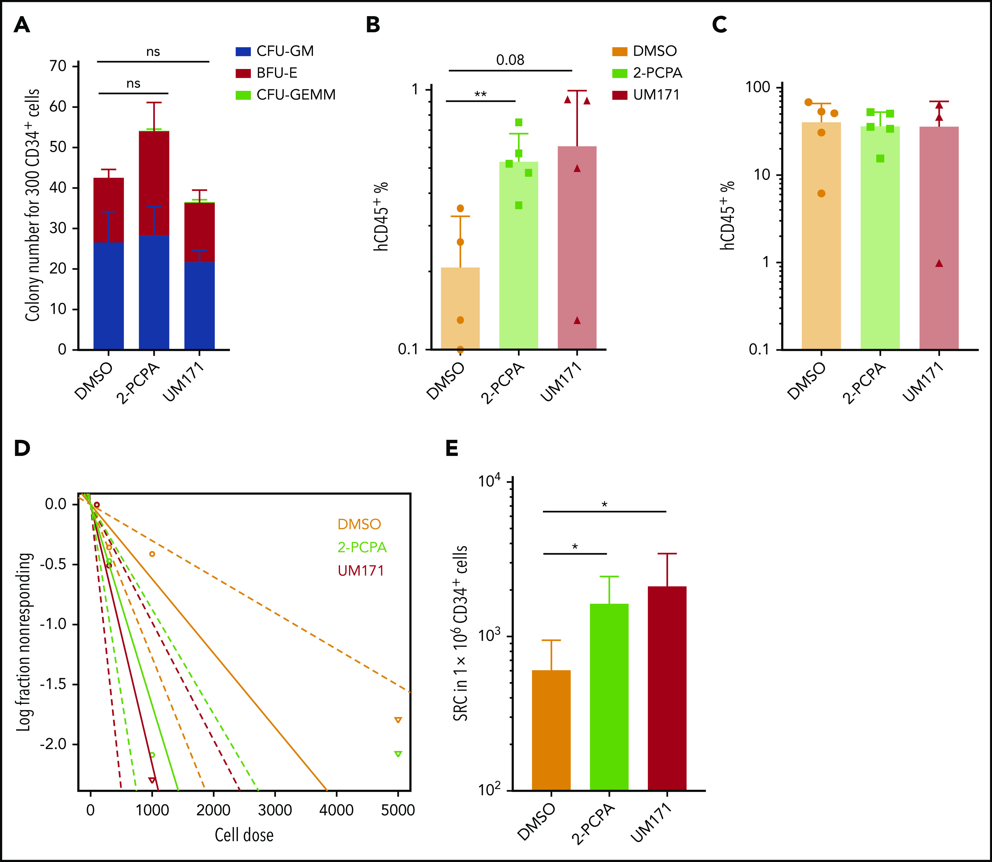
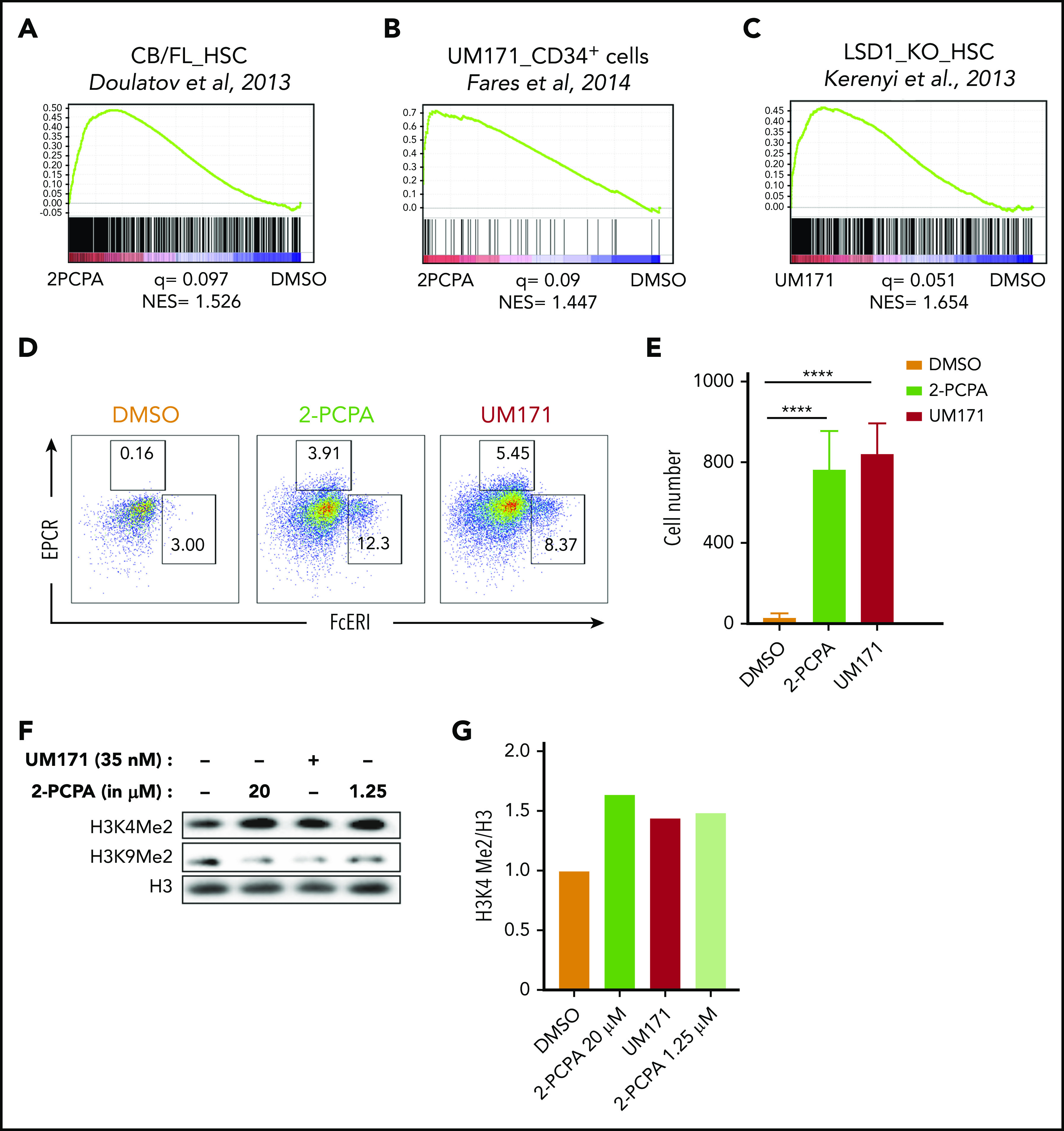
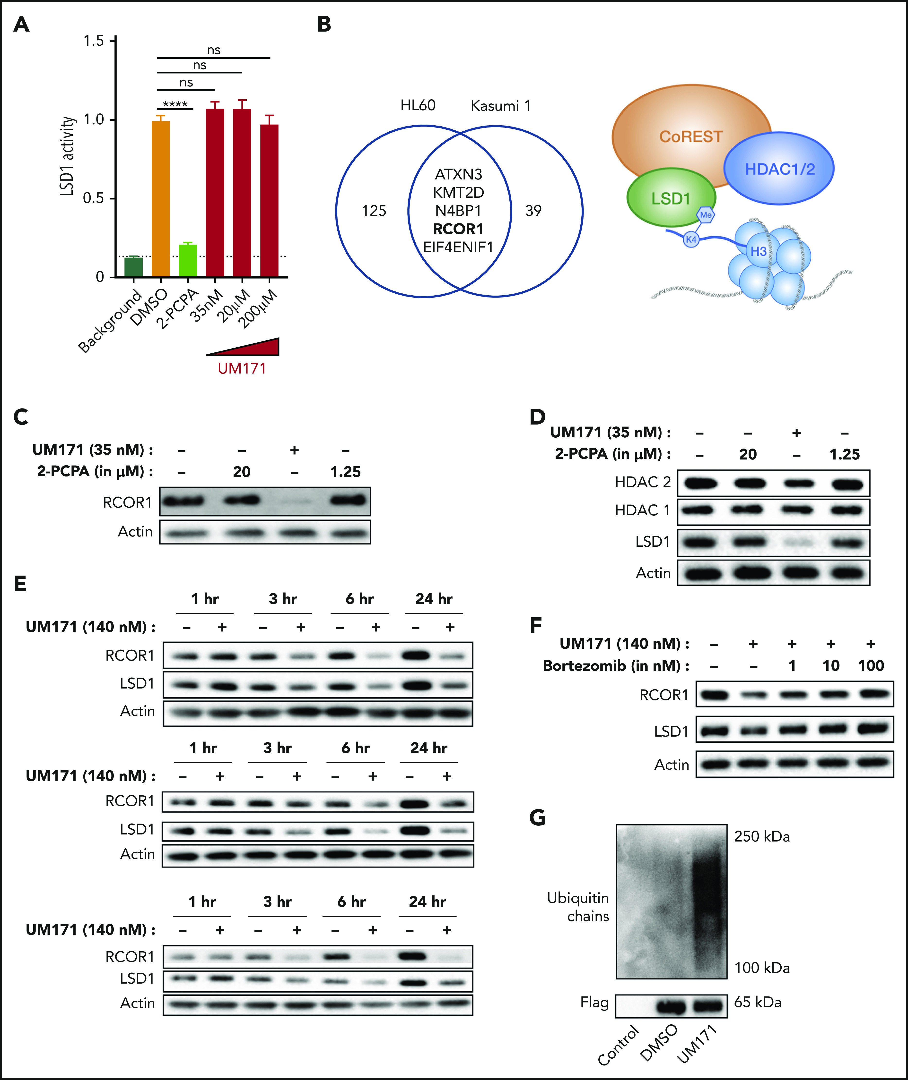
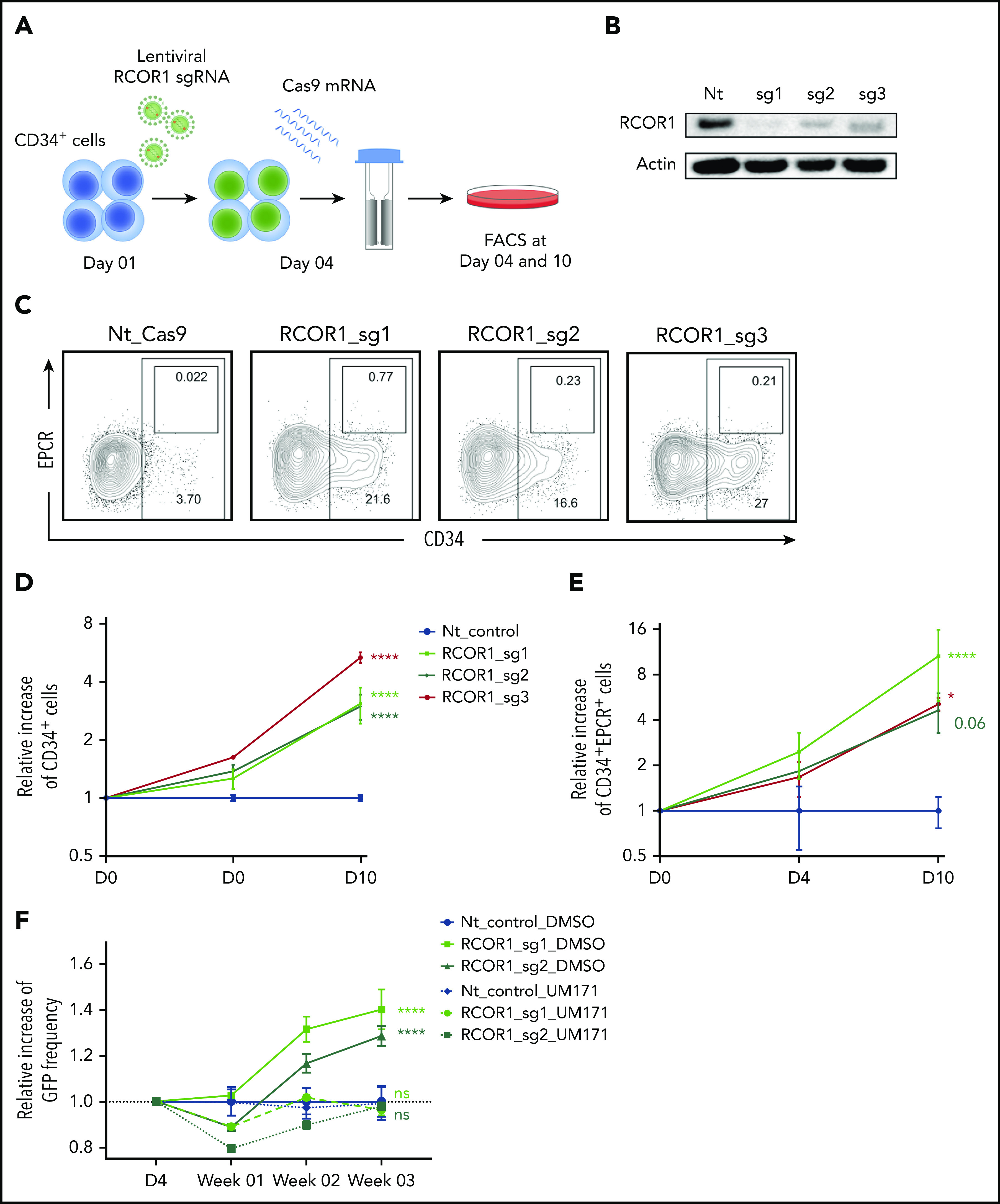
Culture conditions in which hematopoietic stem cells (HSCs) can be expanded for clinical benefit are highly sought after. Here, we report that inhibition of the epigenetic regulator lysine-specific histone demethylase 1A (LSD1) induces a rapid expansion of human cord blood-derived CD34+ cells and promotes in vitro propagation of long-term repopulating HSCs by preventing differentiation. The phenotype and molecular characteristics of cells treated with LSD1 inhibitors were highly similar to cells treated with UM171, an agent promoting expansion of HSCs through undefined mechanisms and currently being tested in clinical trials. Strikingly, we found that LSD1, as well as other members of the LSD1-containing chromatin remodeling complex CoREST, is rapidly polyubiquitinated and degraded upon UM171 treatment. CRISPR (clustered regularly interspaced short palindromic repeats)/Cas9 depletion of the CoREST core member, RCOR1, resulted in expansion of CD34+ cells similar to LSD1 inhibition and UM171. Taken together, LSD1 and CoREST restrict HSC expansion and are principal targets of UM171, forming a mechanistic basis for the HSC-promoting activity of UM171.


Conflict of interest statement

Conflict-of-interest disclosure: The authors declare no competing financial interests.

Figures

None
Graphical abstract
Figure 1.
Figure 1.
LSD1 inhibition promotes expansion of human HSPCs in vitro. (A) Dose-titration experiment showing the effect of 3 LSD1 inhibitors (2-PCPA, GSK-LSD1, or RN-1) on UCB-derived CD34+ HSPC expansion rate compared with the starting cell number (data from 2 repeats). (B) Total numbers of CD34+ cells obtained after treating 10 000 fresh CD34+ cells with 2-PCPA, GSK-LSD1, or RN-1 at their respective half-maximal inhibitory concentration (IC50)/16 concentration (2-PCPA-1.25 μM; GSK_LSD1-1 nM, RN1-4.37 nM) for 6 days. (C) FACS plots showing the frequency (C) and graph showing the total numbers (D) of CD34+EPCR+ cells obtained after treating 10 000 fresh CD34+ cells with 2-PCPA, GSK-LSD1 or RN-1, at their respective IC50/16 concentration, for 6 days. Data from 3 replicates from 1 of 5 independent experiments with similar results are shown. Frequency (E) and numbers (F) of CD34+EPCR+ cells obtained after treating UCB CD34+CD38CD45RACD90+ cells with 2-PCPA for 6 days. (G) Total numbers of CD34+EPCR+ cells obtained after treating BM-derived CD34+CD38CD45RACD90+ cells with 2-PCPA for 6 days. Data from 3 replicates from 1 of 2 independent experiments with similar results. *P ≤ .05, **P ≤ .01, ***P ≤ .001, ****P ≤ .0001. ns, not significant.
Figure 2.
Figure 2.
LSD1 inhibition enhances the ex vivo propagation of long-term repopulating HSCs. (A) Colony-forming capacity of CD34+ cells expanded with 2-PCPA or UM171 for 6 days. Accumulated data from 2 independent experiments with 3 replicates each. hCD45 engraftment in peripheral blood at week 5 (B) and hCD45 engraftment in BM at week 16 (C) in NSG mice transplanted with the progeny of 20 000 plated UCB CD34+ cells expanded with DMSO, 2-PCPA, or UM171 for 6 days. Five mice per each group was used as starting number. (D) Plot summarizing data obtained from LDA analysis. (E) Total number of SRCs obtained from LDA of cultures treated with DMSO, 2-PCPA, or UM171. Numbers are normalized to 1 million CD34+ cells before culture. *P ≤ .05, **P ≤ .01, generalized linear models.
Figure 3.
Figure 3.
LSD1 inhibition and UM171 trigger similar molecular responses. GSEA plots showing HSC gene signature (A) and UM171-induced gene signature (B) in UCB HSC fraction treated with 2-PCPA for 24 hours. (C) LSD1-knockout (KO) gene signature in UCB HSC fraction treated with UM171 for 6 days. Microarray was performed in triplicates, and normalized enrichment score (NES) and q value were calculated with GSEA using reference gene sets from the indicated publications.,, FACS plots showing the frequency (D) and graphs showing the total numbers (E) of mast cell progenitors in cultures treated with 2-PCPA or UM171. Data from 4 replicates from 1 of 2 independent experiments with similar results. (F) Western blot showing H3K4me2 and H3K9me2 marks after 2-PCPA or UM171 treatment. (G) Relative increase in H3K4me2 compared with H3. ****P ≤ .0001.
Figure 4.
Figure 4.
LSD1 and the CoREST complex are targeted by UM171. (A) In vitro assay measuring LSD1 enzymatic activity in the presence of 2-PCPA or UM171. Data from 3 to 6 repeats of the assay are represented. (B) Venn diagram showing common target proteins of UM171 between HL-60 and Kasumi-1 cells, identified by TPP. Western blots showing RCOR1 (C) and LSD1, HDAC1, and HDAC2 (D) in UCB CD34+ cells treated with 2-PCPA or UM171 for 24 hours. (E) Time kinetics experiment showing RCOR1 and LSD1 protein expression in OCI-AML3 (top panel), HL-60 (middle panel), and U937 (bottom panel) cell lines treated with DMSO or UM171. (F) RCOR1 and LSD1 protein expression in HL-60 cell line treated with UM171 and proteasomal inhibitor bortezomib for 3 hours. (G) Flag-tagged RCOR1-expressing HL-60 cells were treated with DMSO or UM171 for 1.5 hours, and RCOR1 was enriched with Flag IP and analyzed for polyubiquitination. ****P ≤ .0001.
Figure 5.
Figure 5.
Loss of CoREST (RCOR1) induces HSPC expansion. (A) Schematic representation of the experimental strategy to knockout RCOR1. (B) RCOR1 protein levels in UCB CD34+ cells edited with nontargeting (Nt) control or RCOR1 guides (day 10). (C) FACS plots showing the frequency of CD34+ and EPCR+ populations in Nt control and RCOR1-knockout UCB HSPCs (day 10). The relative number of CD34+ cells (D) and CD34+EPCR+ cells (E) in RCOR1-knockout HSPCs compared with the Nt control–treated cells over time. Total cell numbers for each condition and time point were calculated and normalized to the mean of the control Nt sgRNA. Data from 3 replicates from 1 of 2 independent experiments with similar results. (F) Relative change in frequency of GFP+ cells over time in Nt control and RCOR1-edited samples compared with day 4 (D4) in DMSO- or UM171-treated cultures. Values are normalized to the NT DMSO control (n = 3). *P ≤ .05, ****P ≤ .0001.

Comment in

Similar articles

Cited by

References

    1. Laughlin MJ, Barker J, Bambach B, et al. . Hematopoietic engraftment and survival in adult recipients of umbilical-cord blood from unrelated donors. N Engl J Med. 2001;344(24):1815-1822. - PubMed
    1. Lund TC, Boitano AE, Delaney CS, Shpall EJ, Wagner JE. Advances in umbilical cord blood manipulation-from niche to bedside. Nat Rev Clin Oncol. 2015;12(3):163-174. - PMC - PubMed
    1. Boitano AE, Wang J, Romeo R, et al. . Aryl hydrocarbon receptor antagonists promote the expansion of human hematopoietic stem cells. Science. 2010;329(5997):1345-1348. - PMC - PubMed
    1. Baudet A, Karlsson C, Safaee Talkhoncheh M, Galeev R, Magnusson M, Larsson J. RNAi screen identifies MAPK14 as a druggable suppressor of human hematopoietic stem cell expansion. Blood. 2012;119(26):6255-6258. - PubMed
    1. Chaurasia P, Gajzer DC, Schaniel C, D’Souza S, Hoffman R. Epigenetic reprogramming induces the expansion of cord blood stem cells. J Clin Invest. 2014;124(6):2378-2395. - PMC - PubMed

Publication types

MeSH terms