. 2022 Mar 15;225(6):1005-1010.
doi: 10.1093/infdis/jiaa367.

A Role for Taok2 in Listeria monocytogenes Vacuolar Escape

Affiliations

A Role for Taok2 in Listeria monocytogenes Vacuolar Escape

Juan J Quereda et al. J Infect Dis. .

Abstract

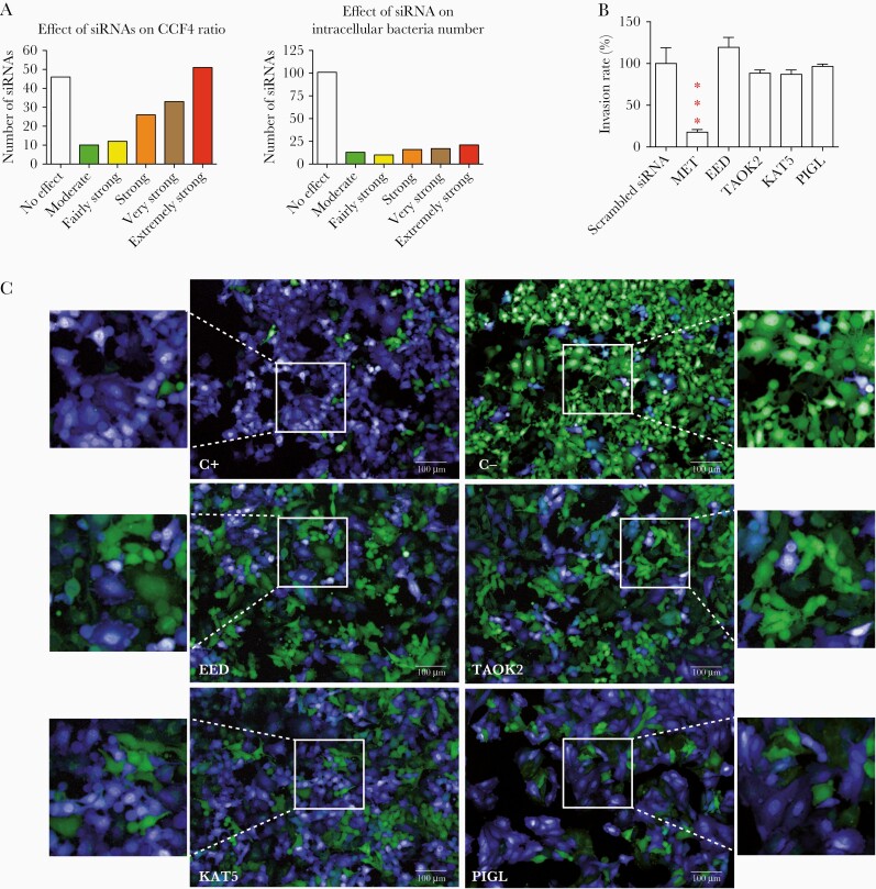
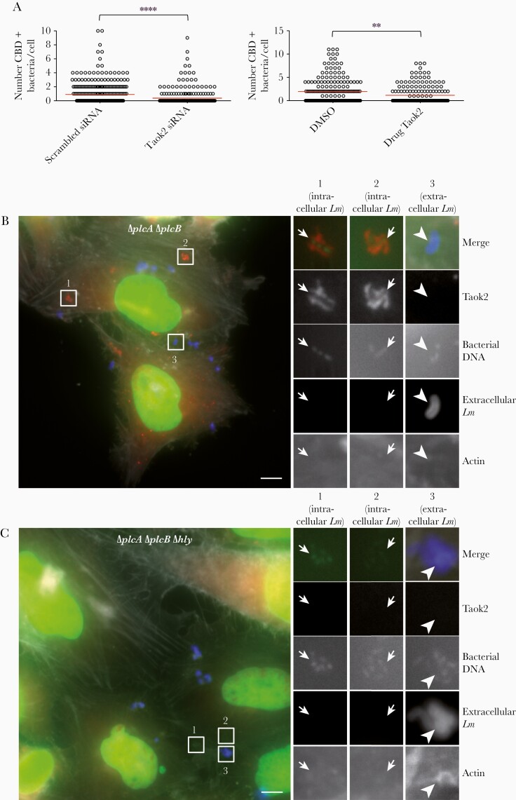
The bacterial pathogen Listeria monocytogenes invades host cells, ruptures the internalization vacuole, and reaches the cytosol for replication. A high-content small interfering RNA (siRNA) microscopy screen allowed us to identify epithelial cell factors involved in L. monocytogenes vacuolar rupture, including the serine/threonine kinase Taok2. Kinase activity inhibition using a specific drug validated a role for Taok2 in favoring L. monocytogenes cytoplasmic access. Furthermore, we showed that Taok2 recruitment to L. monocytogenes vacuoles requires the presence of pore-forming toxin listeriolysin O. Overall, our study identified the first set of host factors modulating L. monocytogenes vacuolar rupture and cytoplasmic access in epithelial cells.

Keywords: Listeria monocytogenes; STE1-like kinase; Taok2; siRNA screen; vacuolar escape.


Figures

Figure 1.
Figure 1.
High-content microscopy siRNA screen reveals host factors required by Listeria monocytogenes for vacuolar escape in epithelial cells. A, Overview of the screen results. After data normalization based on the strictly standardized mean difference method, 165 targeted genes were classified into 6 categories based on their effect on the FRET probe CCF4 green to blue ratio (vacuolar escape step, left) and in the intracellular bacteria number (entry step, right): no effect, moderate, fairly strong, strong, very strong, and extremely strong effects. B, Contributions of MET (control for invasion), EED, TAOK2, KAT5, and PIGL to L. monocytogenes invasion. Target genes were knocked down by siRNA transfection of HeLa cells. One hour after infection cells were washed and L. monocytogenes entry was measured by counting colony forming units. Bars represent the mean and SD from 3 experiments. Significant differences from the scramble treatment *** P < .001. C, Examples of images from the siRNA screen. HeLa cells were grown in 96-well plates and transfected by siRNAs. Host cells were loaded with CCF4-AM and infected with L. monocytogenes EGDe PrfA* β-lact (C+) or L. innocuaβ-lact/InlB (C−) for 1 hour. C+ and C− negative wells were transfected with scrambled siRNA. After paraformaldehyde fixation, nuclei were stained with Draq5 and cells were imaged using an Opera QEHS confocal microscope, as previously described [3]. Images were obtained with the following merged channels: the intact CCF4 probe peaks at 535 nm (green), and the cleaved CCF4 probe peaks at 450 nm (blue). L. monocytogenes EGDe PrfA* β-lact (C+) can escape its vacuolar compartments and induce the cleavage of the CCF4 probe (top left). Scale bar 100µm. Abbreviations: FRET, fluorescence resonance energy transfer; siRNA, small interfering RNA.
Figure 2.
Figure 2.
Taok2 siRNA knockdown or inhibition of its kinase activity by the chemical compound SW172006 impairs efficient rupture of Listeria monocytogenes-containing vacuoles. A, Taok2 knockdown by siRNA (left) or inhibition of its kinase activity (right) results in reduced cytoplasmic access. HeLa cells were transfected with the YFP-CBD of the L. monocytogenes phage endolysin Ply118, which identifies cytosolic bacteria shortly after escape. The red line indicates the mean. Results were from 100 cells counted in 3 representative fields to estimate the number of CBD-labeled bacteria. Statistical analysis: 2-tailed unpaired t tests. **P < .01, ****P < .0001. B and C, Taok2 accumulation in a compartment resembling a L. monocytogenes-containing vacuole. B, HeLa cells infected with L. monocytogenes EGDe PrfA*ΔplcAΔplcB for 30 minutes and imaged. Taok2 (labeled with anti-Taok2 antibodies) is shown in red, DNA (labeled with Hoechst) is shown in green, extracellular bacteria (labeled with anti-L. monocytogenes antibodies) are shown in blue, and actin (labeled with phalloidin) is shown in white. Left image: overview. Right images: enlargement of 3 ROIs shown in the overview. In ROIs 1 and 2, Taok2 decorates intracellular bacteria (small arrows, revealed by the bacterial DNA labeling and by the absence of extracellular bacterial labeling), which are present in a vacuolar compartment and do not polymerize cytoplasmic actin (absence of actin labeling). In ROI 3, extracellular bacteria (large arrowhead, revealed by the presence of both DNA and extracellular L. monocytogenes labeling) are not decorated by Taok2. Scale bar, 5 μm. C, HeLa cells infected as in (B) using the strain L. monocytogenes EGDe PrfA* ΔplcAΔplcBΔhly. Left image: overview. Right images: enlargement of 3 ROIs shown in the overview. In all the ROIs showing either intracellular or extracellular L. monocytogenes Taok2 does not decorate the bacteria. Scale bar, 5 μm. Abbreviations: CBD, cell wall binding domain; DMSO, dimethyl sulfoxide; LM, Listeria monocytogenes; ROI, regions of interest; siRNA, small interfering RNA; YFP, yellow fluorescent protein.

References

    1. Pizarro-Cerdá J, Cossart P. Microbe profile: Listeria monocytogenes: a paradigm among intracellular bacterial pathogens. Microbiology 2019; 165:719–21. - PubMed
    1. Pizarro-Cerda J, Cossart P. Listeria monocytogenes: cell biology of invasion and intracellular growth. Microbiol Spectr 2018; 6: 10.1128/microbiolspec.GPP3-0013-2018. - PMC - PubMed
    1. Quereda JJ, Pizarro-Cerdá J, Balestrino D, et al. . A dual microscopy-based assay to assess Listeria monocytogenes cellular entry and vacuolar escape. Appl Environ Microbiol 2016; 82:211–7. - PMC - PubMed
    1. Mellouk N, Weiner A, Aulner N, et al. . Shigella subverts the host recycling compartment to rupture its vacuole. Cell Host Microbe 2014; 16:517–30. - PubMed
    1. Weiss DS, Brotcke A, Henry T, Margolis JJ, Chan K, Monack DM. In vivo negative selection screen identifies genes required for Francisella virulence. Proc Natl Acad Sci U S A 2007; 104:6037–42. - PMC - PubMed

Publication types