. 2020 Sep;46(3):1013-1028.
doi: 10.3892/ijmm.2020.4649. Epub 2020 Jun 18.

MicroRNA‑103 modulates tumor progression by targeting KLF7 in non‑small cell lung cancer

Affiliations

MicroRNA‑103 modulates tumor progression by targeting KLF7 in non‑small cell lung cancer

Ke Li et al. Int J Mol Med. 2020 Sep.

Abstract

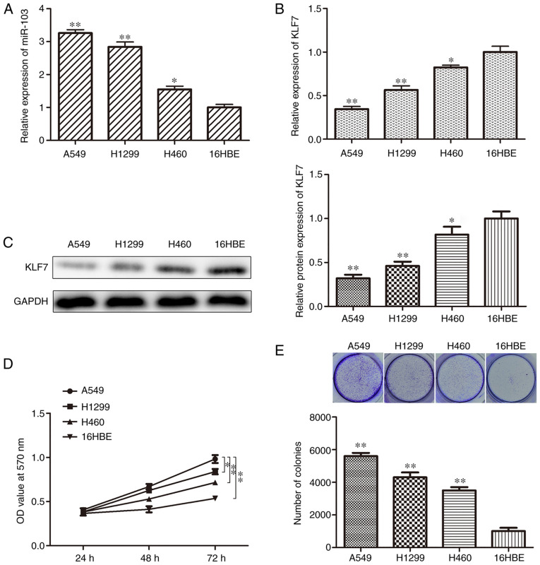
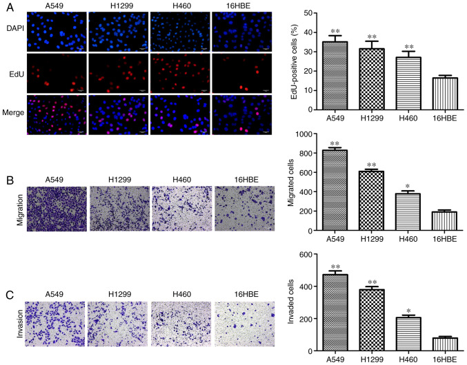
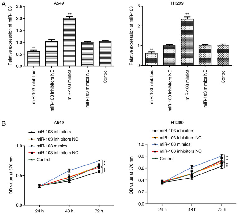
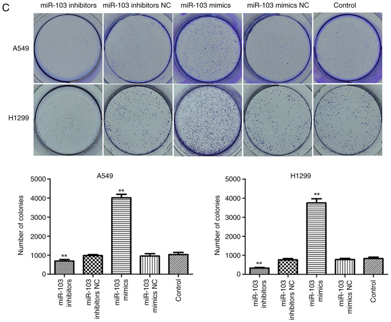
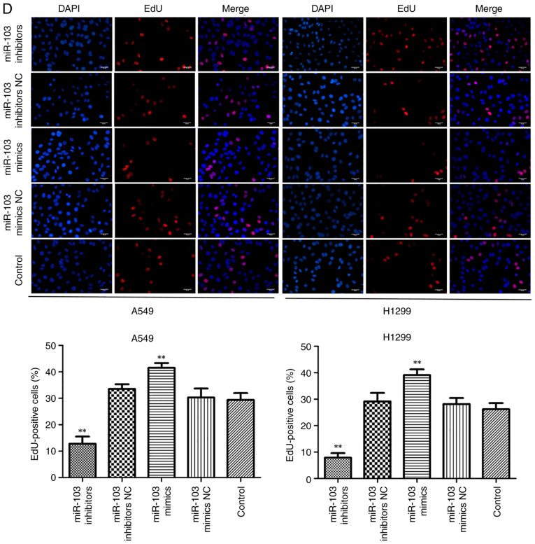
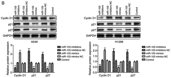
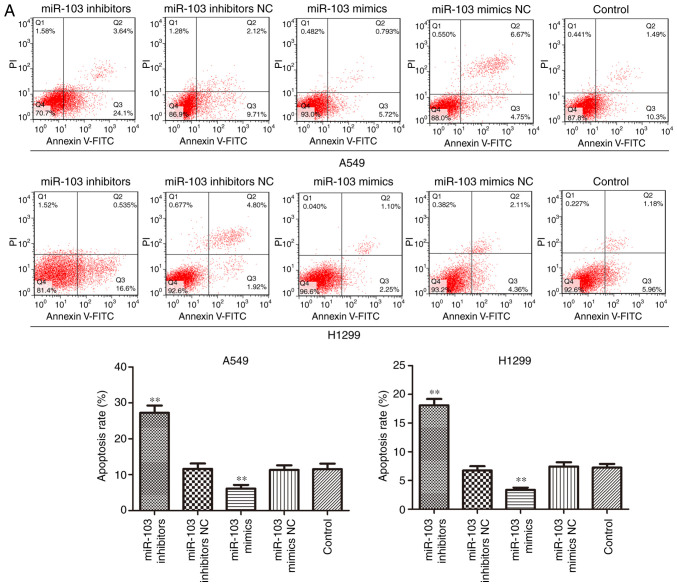
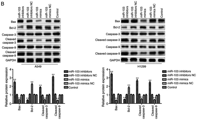
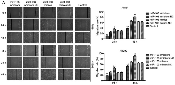
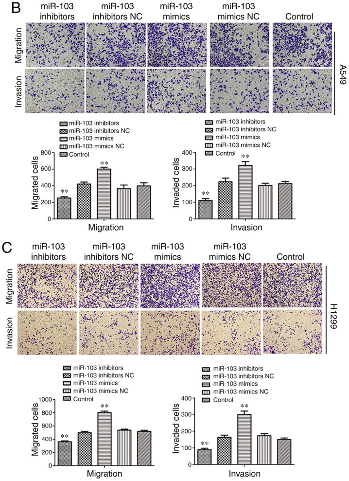
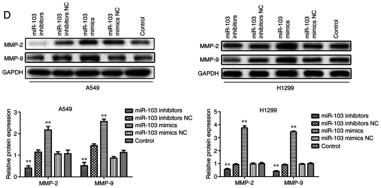
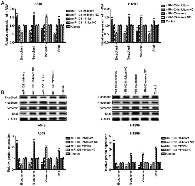
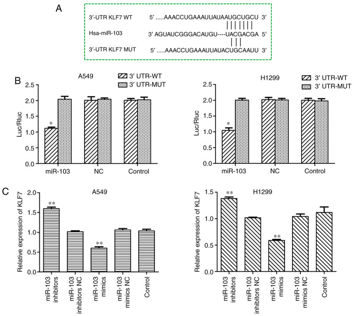
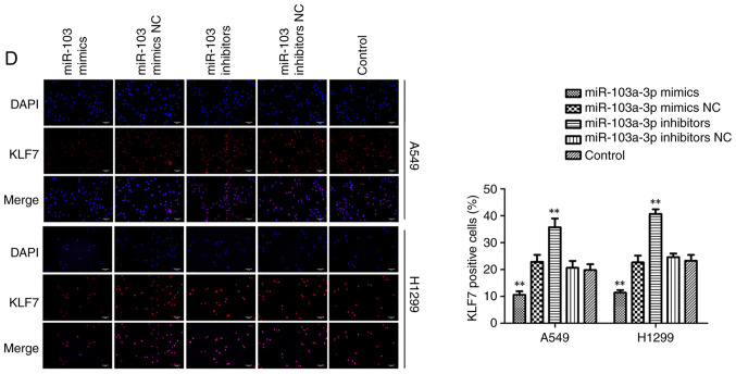
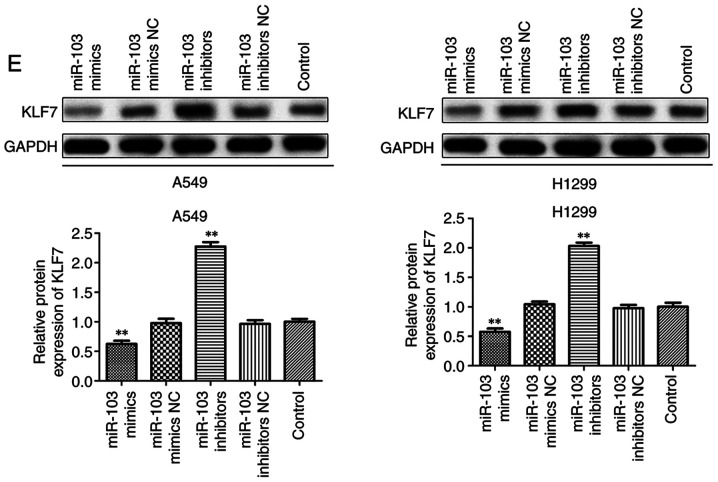
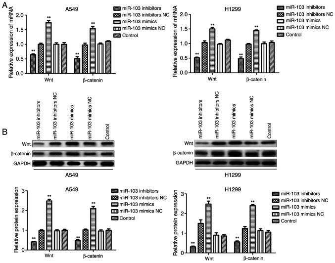
Numerous studies have identified that microRNAs (miRs) play a crucial role in the tumorigenesis of non‑small cell lung cancer (NSCLC). However, to the best of our knowledge, the physiological function of miR‑103 in NSCLC is not fully understood. Experiments in the present study revealed that miR‑103 expression was increased in NSCLC cell lines. In addition, a series of methods, including MTT, colony formation, 5‑ethynyl‑2'‑deoxyuridine, Transwell, wound healing, flow cytometric, reverse transcription‑quantitative PCR and western blot assays, were performed, which revealed that overexpression of miR‑103 enhanced cell growth, migration, invasion and epithelial‑mesenchymal transition (EMT), and suppressed apoptosis of A549 and H1299 cells. Additionally, a dual‑luciferase reporter assay indicated that miR‑103 directly targets the 3'‑untranslated region of Kruppel‑like factor 7 (KLF7), and KLF7 expression was negatively regulated by miR‑103 expression. Furthermore, the present findings demonstrated that miR‑103 promoted EMT via regulating the Wnt/β‑catenin signaling pathway in NSCLC. Collectively, the current results demonstrated that miR‑103 serves a tumorigenesis role in NSCLC development by targeting KLF7, at least partly via the Wnt/β‑catenin signaling pathway. Consequently, these findings indicated that miR‑103/KLF7/Wnt/β‑catenin may provide a novel insight into underlying biomarkers for improving the diagnosis and treatment of NSCLC.


Figures

Figure 1
Figure 1
miR-103 is highly expressed in NSCLC cell lines. (A) The expression levels of miR-103 in NSCLC cell lines and the 16HBE cell line were examined by RT-qPCR. *P<0.05, **P<0.01 vs. 16HBE cells. The expression of KLF7 was detected by (B) RT-qPCR and (C) western blot assay in NSCLC cells and 16HBE cells. (D) Proliferation and (E) colony formation ability of cells were determined by MTT and colony formation assays, respectively. Data are presented as the mean ± standard deviation (n=3). *P<0.05, **P<0.01 vs. 16HBE cells. miR-103, microRNA-103; NSCLC, non-small cell lung cancer; RT-qPCR, reverse transcription-quantitative PCR; KLF7, Kruppel-like factor 7; OD, optical density.
Figure 2
Figure 2
Proliferation, migration and invasion of NSCLC cell lines. (A) Proliferation of cells was determined by EdU assay. Magnification, ×400. Scale bar, 50 µm. The (B) migration and (C) invasion of NSCLC cells and 16HBE cells were detected by Transwell assays. Magnification, ×100. Scale bar, 100 µm. *P<0.05, **P<0.01 vs. 16HBE cells. NSCLC, non-small cell lung cancer; EdU, 5-ethynyl-2′-deoxyuridine.
Figure 3
Figure 3
miR-103 promotes cell viability in non-small cell lung cancer. (A) miR-103 expression levels in cell lines transfected with miR-103 mimic and miR-103 inhibitor were measured by reverse transcription-quantitative PCR assay. **P<0.01 vs. control. (B) Cell proliferation was assessed by a MTT assay. **P<0.01. (C) Colony formation assay was performed with transfected A549 and H1299 cells. Magnification, ×400. miR-103 promotes cell viability in non-small cell lung cancer. (D) EdU assay was performed with transfected A549 and H1299 cells. Scale bar, 50 µm. **P<0.01 vs. control. miR-103, microRNA-103; NC, negative control; EdU, 5-ethynyl-2′-deoxyuridine; OD, optical density.
Figure 3
Figure 3
miR-103 promotes cell viability in non-small cell lung cancer. (A) miR-103 expression levels in cell lines transfected with miR-103 mimic and miR-103 inhibitor were measured by reverse transcription-quantitative PCR assay. **P<0.01 vs. control. (B) Cell proliferation was assessed by a MTT assay. **P<0.01. (C) Colony formation assay was performed with transfected A549 and H1299 cells. Magnification, ×400. miR-103 promotes cell viability in non-small cell lung cancer. (D) EdU assay was performed with transfected A549 and H1299 cells. Scale bar, 50 µm. **P<0.01 vs. control. miR-103, microRNA-103; NC, negative control; EdU, 5-ethynyl-2′-deoxyuridine; OD, optical density.
Figure 4
Figure 4
Effect of miR-103 on cell cycle in non-small cell lung cancer. (A) Cell cycle analysis was performed by flow cytometry to determine the impact of miR-103 on cell cycle progression. Overexpression of miR-103 inhibited cell cycle progression, whereas knockdown of miR-103 promoted cell cycle progression. (B) The cell cycle-related proteins p21, p27 and cyclin D1 were measured in A549 and H1299 cells using western blot assays. Data are presented as the mean ± standard deviation (n=3). **P<0.01 vs. control. miR-103, microRNA-103; NC, negative control.
Figure 4
Figure 4
Effect of miR-103 on cell cycle in non-small cell lung cancer. (A) Cell cycle analysis was performed by flow cytometry to determine the impact of miR-103 on cell cycle progression. Overexpression of miR-103 inhibited cell cycle progression, whereas knockdown of miR-103 promoted cell cycle progression. (B) The cell cycle-related proteins p21, p27 and cyclin D1 were measured in A549 and H1299 cells using western blot assays. Data are presented as the mean ± standard deviation (n=3). **P<0.01 vs. control. miR-103, microRNA-103; NC, negative control.
Figure 5
Figure 5
Effect of miR-103 on cell apoptosis in non-small cell lung cancer. (A) Cell apoptosis was analyzed by flow cytometry of A549 and H1299 cells after transfection with miR-103 mimics, miR-103 inhibitors and the NC. (B) Apoptotic proteins were analyzed using western blotting. Data are presented as the mean ± standard deviation (n=3). **P<0.01 vs. control. miR-103, microRNA-103; NC, negative control; PI, propidium iodide.
Figure 5
Figure 5
Effect of miR-103 on cell apoptosis in non-small cell lung cancer. (A) Cell apoptosis was analyzed by flow cytometry of A549 and H1299 cells after transfection with miR-103 mimics, miR-103 inhibitors and the NC. (B) Apoptotic proteins were analyzed using western blotting. Data are presented as the mean ± standard deviation (n=3). **P<0.01 vs. control. miR-103, microRNA-103; NC, negative control; PI, propidium iodide.
Figure 6
Figure 6
miR-103 promotes cell migration and invasion in NSCLC cells. The migration and invasion of transfected cells were investigated by (A) wound healing and Transwell assays with (B) A549 and (C) H1299 cells. Magnification ×100. miR-103 promotes cell migration and invasion in NSCLC cells. (D) The protein expression levels of MMP-2 and MMP-9 in A549 and H1299 cells were measured by western blotting. *P<0.05 and **P<0.01 vs. control. miR-103, microRNA-103; NC, negative control; MMP, matrix metallopeptidase.
Figure 6
Figure 6
miR-103 promotes cell migration and invasion in NSCLC cells. The migration and invasion of transfected cells were investigated by (A) wound healing and Transwell assays with (B) A549 and (C) H1299 cells. Magnification ×100. miR-103 promotes cell migration and invasion in NSCLC cells. (D) The protein expression levels of MMP-2 and MMP-9 in A549 and H1299 cells were measured by western blotting. *P<0.05 and **P<0.01 vs. control. miR-103, microRNA-103; NC, negative control; MMP, matrix metallopeptidase.
Figure 7
Figure 7
miR-103 promotes the epithelial-mesenchymal transition of non-small cell lung cancer cells. The expression levels of E-cadherin, N-cadherin, Vimentin and Snail in A549 and H1299 cells were measured by (A) reverse transcription-quantitative PCR and (B) western blot analysis. GAPDH served as a loading control. **P<0.01 vs. control. miR-103, microRNA-103; NC, negative control.
Figure 8
Figure 8
miR-103 suppresses the expression of KLF7 in NSCLC cells by binding to the 3′-UTR of the KLF7 gene. (A) miR-103 and its putative binding sequence in the 3′-UTR of KLF7. (B) A dual-luciferase reporter assay was performed to further confirm whether miR-103 can directly target the 3′-UTR region of KLF7 in NSCLC cells. *P<0.05 vs. 3′-UTR-MUT. The expression levels of KLF7 in A549 and H1299 cells transfected with miR-103 mimic or inhibitor were measured by (C) reverse transcription-quantitative PCR and (D) western blot analysis. **P<0.01 vs. control. miR-103 suppresses the expression of KLF7 in NSCLC cells by binding to the 3′-UTR of the KLF7 gene. (E) The expression of KLF7 regulated by miR-103 in A549 and H1299 cells was assessed by immunofluorescence analysis. Magnification, ×200. Scale bar, 50 µm. **P<0.01 vs. control. miR-103, microRNA-103; NC, negative control; KLF7, Kruppel-like factor 7; 3′-UTR, 3′-untranslated region; NSCLC, non-small cell lung cancer; Luc, luciferase; Rluc, Renilla luciferase; WT, wild-type; MUT, mutant.
Figure 8
Figure 8
miR-103 suppresses the expression of KLF7 in NSCLC cells by binding to the 3′-UTR of the KLF7 gene. (A) miR-103 and its putative binding sequence in the 3′-UTR of KLF7. (B) A dual-luciferase reporter assay was performed to further confirm whether miR-103 can directly target the 3′-UTR region of KLF7 in NSCLC cells. *P<0.05 vs. 3′-UTR-MUT. The expression levels of KLF7 in A549 and H1299 cells transfected with miR-103 mimic or inhibitor were measured by (C) reverse transcription-quantitative PCR and (D) western blot analysis. **P<0.01 vs. control. miR-103 suppresses the expression of KLF7 in NSCLC cells by binding to the 3′-UTR of the KLF7 gene. (E) The expression of KLF7 regulated by miR-103 in A549 and H1299 cells was assessed by immunofluorescence analysis. Magnification, ×200. Scale bar, 50 µm. **P<0.01 vs. control. miR-103, microRNA-103; NC, negative control; KLF7, Kruppel-like factor 7; 3′-UTR, 3′-untranslated region; NSCLC, non-small cell lung cancer; Luc, luciferase; Rluc, Renilla luciferase; WT, wild-type; MUT, mutant.
Figure 9
Figure 9
miR-103 activates the Wnt/β-catenin signaling pathway in non-small cell lung cancer cells. The (A) mRNA and (B) protein expression levels of Wnt and β-catenin were determined in transfected A549 and H1299 cells. Data are presented as the mean ± standard deviation. **P<0.01 vs. control. miR-103, microRNA-103; NC, negative control.

Similar articles

Cited by

References

    1. Bray F, Ferlay J, Soerjomataram I, Siegel RL, Torre LA, Jemal A. Global cancer statistics 2018: GLOBOCAN estimates of incidence and mortality worldwide for 36 cancers in 185 countries. CA Cancer J Clin. 2018;68:394–424. doi: 10.3322/caac.21492. - DOI - PubMed
    1. Duma NR, Santana-Davila, Molina JR. Non-small cell lung cancer: Epidemiology, screening, diagnosis, and treatment. Mayo Clin Proc. 2019;94:1623–1640. doi: 10.1016/j.mayocp.2019.01.013. - DOI - PubMed
    1. Ettinger DS, Wood DE, Akerley W, Bazhenova LA, Borghaei H, Camidge DR, Cheney RT, Chirieac LR, D'Amico TA, Demmy TL, et al. Non-small cell lung cancer, version 1.2015. J Natl Compr Canc Netw. 2014;12:1738–1761. doi: 10.6004/jnccn.2014.0176. - DOI - PubMed
    1. Gompelmann D, Eberhardt R, Herth FJ. Advanced malignant lung disease: What the specialist can offer. Respiration. 2011;82:111–123. doi: 10.1159/000329703. - DOI - PubMed
    1. Goldstraw P, Chansky K, Crowley J, Porta RR, Asamura H, Eberhardt WE, Nicholson AG, Groome P, Mitchell A, Bolejack V, et al. The IASLC lung cancer staging project: Proposals for revision of the tnm stage groupings in the forthcoming (Eighth) edition of the TNM classification for lung cancer. J Thorac Oncol. 2016;11:39–51. doi: 10.1016/j.jtho.2015.09.009. - DOI - PubMed

MeSH terms