. 2020 Sep;22(3):1859-1867.
doi: 10.3892/mmr.2020.11260. Epub 2020 Jun 19.

Age‑related changes in mineralocorticoid receptors in rat hearts

Affiliations

Age‑related changes in mineralocorticoid receptors in rat hearts

Danli Hu et al. Mol Med Rep. 2020 Sep.

Abstract

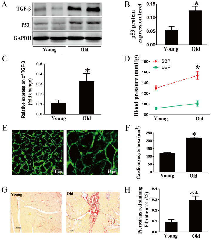
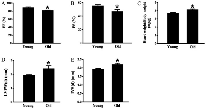
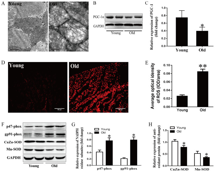
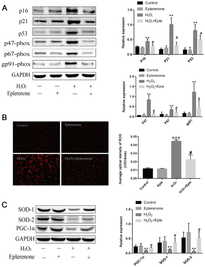
Age-related alterations in the renin-angiotensin-aldosterone system (RAAS) have been reported in the cardiovascular system; however, the detailed mechanism of the RAAS component mineralocorticoid receptors (MR) has not been elucidated. The present study aimed to investigate the associations between MR and cardiac aging in rats, as well as the regulatory effects of oxidative stress and mitochondrial abnormalities in the aging process. MR expression in the hearts of male Sprague‑Dawley rats aged 3 months (young rats) and 24 months (old rats) was evaluated in vivo. In addition, in vitro, H9C2 cells were treated with a specific MR antagonist, eplerenone, in order to investigate the molecular mechanism underlying the inhibition of myocyte aging process. The results demonstrated that MR expression was significantly higher in 24‑month‑old rat hearts compared with in 3‑month‑old rat hearts. These changes were accompanied by increased p53 expression, decreased peroxisome proliferator‑activated receptor γ coactivator‑1α expression, decreased mitochondrial renewal as assessed by electron microscopy, increased oxidative stress and decreased superoxide dismutase. In vitro, selective antagonism of MR partially blocked H2O2‑induced myocardial aging as assessed by p16, p21 and p53 expression levels and excessive reactive oxygen species (ROS) accumulation. These results indicated that increased MR expression may drive age‑related cardiac dysfunction via mitochondrial damage, increased ROS accumulation and an imbalanced redox state.

Keywords: mineralocorticoid receptors; cardiac aging; oxidative stress; mitochondrial dysfunction; redox state.


Figures

Figure 1.
Figure 1.
Effects of aging on myocardial hypertrophy and fibrosis. (A) Representative immunoblots, and (B) semi-quantification for p53 and (C) TGF-β1 in cardiac tissues. (D) SBP and DBP of 3- and 24-month-old rats. (E) Cardiomyocyte area determined by WGA staining and (F) the corresponding semi-quantification of WGA staining; scale bar, 15 µm. (G) Myocardial interstitial and perivascular fibrosis stained by Picrosirius red and (H) the corresponding semi-quantification; scale bar, 100 µm. Data are presented as the means ± standard error of the mean from each group; n=8 per group. *P<0.05, **P<0.01 vs. young group. DBP, diastolic blood pressure; SBP, systolic blood pressure; TGF-β1, transforming growth factor-β1.
Figure 2.
Figure 2.
Effects of aging on cardiac function. (A) EF and (B) FS were measured by echocardiography. (C) Ratio of heart weight to body weight was detected after the rats were sacrificed. (D) LVPW-d and (E) IVS-d were determined by echocardiography. Data are presented as the means ± standard error of the mean from each group; n=8 per group. *P<0.05 vs. young group. EF, ejection fraction; FS, fractional shortening; IVS-d, interventricular septal thickness at end-diastole; LVPW-d, left ventricular posterior wall thickness at end-diastole.
Figure 3.
Figure 3.
Increased MR expression in the aging heart. (A) Reverse transcription-quantitative PCR analysis of MR mRNA expression in 3- and 24-month-old rats. (B) Representative immunoblots and (C) semi-quantification of MR in the hearts of the two groups. (D) Immunohistochemical staining and (E) corresponding semi-quantification of MR expression in the hearts of young (3 months) and old (24 months) rats; scale bar, 100 µm. Data are presented as the means ± standard error of the mean from each group; n=8 per group. *P<0.05, **P<0.01 vs. young group. IOD, integrated optical density; MR, mineralocorticoid receptors.
Figure 4.
Figure 4.
Effects of aging on mitochondrial stress and the redox state. (A) Mitochondrial ultrastructure of myocytes detected by electron microscopy; scale bar, 0.5 µm. (B) Representative immunoblots and (C) semi-quantitation of PGC-1α. (D) ROS levels detected by DHE fluorescence staining and (E) corresponding semi-quantification; scale bar, 50 µm. (F) Representative immunoblots of p47-phox, gp91-phox, CuZn- and Mn-SOD. (G) Semi-quantification of p47-phox and gp91-phox. (H) Semi-quantification of CuZn-SOD and Mn-SOD. Data are presented as the means ± standard error of the mean from each group; n=8 per group. *P<0.05, **P<0.01 vs. young group. DHE, dihydroethidium; NADPH, nicotinamide adenine dinucleotide phosphate; PGC-1α, peroxisome proliferator-activated receptor γ coactivator-1α; ROS, reactive oxygen species; SOD, superoxide dismutase.
Figure 5.
Figure 5.
MR antagonism suppresses H2O2-induced cardiac aging and mitochondrial dysfunction. (A) Immunoblotting and semi-quantification of p16, p21, p53 and NADP subunits, p47-phox, p67-phox and gp91-phox expression in H9C2 cells. Protein expression levels were normalized to GAPDH. (B) ROS levels detected by DHE staining in H9C2 cells following different treatments (magnification, ×100; scale bar, 100 µm). (C) SOD-1, SOD-2 and PGC-1α protein expression levels detected by western blotting. Data are representative of three experiments, n=3. **P<0.01 and ***P<0.001 vs. control group; #P<0.05 vs. H2O2 group. DHE, dihydroethidium; MR, mineralocorticoid receptors; PGC-1α, peroxisome proliferator-activated receptor γ coactivator-1α; ROS, reactive oxygen species; SOD, superoxide dismutase; Eple, eplerenone.

References

    1. United Nations, Department of Economic, Social Affairs, Population Division. World Population Ageing 2017 - Highlights (ST/ESA/SER.A/397). United Nations, New York, NY, 2017. https://www.un.org/en/development/desa/population/publications/pdf/agein...
    1. Wyss-Coray T. Ageing, neurodegeneration and brain rejuvenation. Nature. 2016;539:180–186. doi: 10.1038/nature20411. - DOI - PMC - PubMed
    1. Rickard AJ, Morgan J, Bienvenu LA, Fletcher EK, Cranston GA, Shen JZ, Reichelt ME, Delbridge LM, Young MJ. Cardiomyocyte mineralocorticoid receptors are essential for deoxycorticosterone/salt-mediated inflammation and cardiac fibrosis. Hypertension. 2012;60:1443–1450. doi: 10.1161/HYPERTENSIONAHA.112.203158. - DOI - PubMed
    1. Lother A, Berger S, Gilsbach R, Rösner S, Ecke A, Barreto F, Bauersachs J, Schütz G, Hein L. Ablation of mineralocorticoid receptors in myocytes but not in fibroblasts preserves cardiac function. Hypertension. 2011;57:746–754. doi: 10.1161/HYPERTENSIONAHA.110.163287. - DOI - PubMed
    1. López-Otin C, Blasco MA, Partridge L, Serrano M, Kroemer G. The hallmarks of aging. Cell. 2013;153:1194–1217. doi: 10.1016/j.cell.2013.05.039. - DOI - PMC - PubMed