. 2020 Sep;22(3):2063-2075.
doi: 10.3892/mmr.2020.11253. Epub 2020 Jun 18.

Expression levels of the α7 nicotinic acetylcholine receptor in the brains of patients with Alzheimer's disease and their effect on synaptic proteins in SH-SY5Y cells

Affiliations

Expression levels of the α7 nicotinic acetylcholine receptor in the brains of patients with Alzheimer's disease and their effect on synaptic proteins in SH-SY5Y cells

Jia-Mou Ren et al. Mol Med Rep. 2020 Sep.

Abstract

Alzheimer's disease (AD) is a chronic neurodegenerative, and abnormal aggregation of the neurotoxic β amyloid (Aβ) peptide is an early event in AD. The present study aimed to determine the correlation between the nicotinic acetylcholine receptor α7 subunit (α7 nAChR) and Aβ in the brains of patients with AD, and to investigate whether the increased expression levels of the α7 nAChR could alter the neurotoxicity of Aβ. The expression levels of α7 nAChR and Aβ in the brains of patients with AD and healthy brains were analyzed using immunofluorescence. Moreover, SH‑SY5Y cells were used to stably overexpress or silence α7 nAChR expression levels, prior to the treatment with or without 1 µmol/l Aβ1‑42 oligomer (AβO). The mRNA and protein expression levels of α7 nAChR, synaptophysin (SYP), postsynaptic density of 95 kDa (PSD‑95) and synaptosomal‑associated protein of 25 kDa (SNAP‑25) were subsequently analyzed using reverse transcription‑quantitative PCR and western blotting. In addition, the concentration of acetylcholine (ACh) and the activity of acetylcholinesterase (AChE) were analyzed using spectrophotometry, while the cell apoptotic rate was determined using flow cytometry. The expression of Aβ in the brains of patients with AD was found to be significantly increased, whereas the expression of α7 nAChR was significantly decreased compared with the healthy control group. In vitro, the expression levels of α7 nAChR were significantly increased or decreased following the overexpression or silencing of the gene, respectively. Consistent with these observations, the mRNA and protein expression levels of SYP, PSD‑95 and SNAP‑25 were also significantly increased following the overexpression of α7 nAChR and decreased following the genetic silencing of the receptor. In untransfected or negative control cells, the expression levels of these factors and the apoptotic rate were significantly reduced following the exposure to AβO, which was found to be attenuated by α7 nAChR overexpression, but potentiated by α7 nAChR RNA silencing. However, no significant differences were observed in either the ACh concentration or AChE activity following transfection. Collectively, these findings suggested that α7 nAChR may protect the brains of patients with AD against Aβ, as α7 nAChR overexpression increased the expression levels of SYP, SNAP‑25 and PSD‑95, and attenuated the inhibitory effect of Aβ on the expression of these synaptic proteins and cell apoptosis. Overall, this indicated that α7 nAChR may serve an important neuroprotective role in AD.

Keywords: SH-SY5Y cells; α7 nicotinic acetylcholine receptor; β-amyloid peptide; synaptic protein; synaptophysin; apoptotic rate; synaptosomal-associated protein of 25 kda; postsynaptic density of 95 kda; alzheimer's disease.


Figures

Figure 1.
Figure 1.
Expression levels of α7 nAChR and Aβ in the brains of patients with AD and normal brains. (A) Images of the immunofluorescence double staining in the control and AD groups. (B) Aβ is expressed in the cytoplasm and extracellular matrix of cells. It was also found highly expressed in the brain slices of patients with AD, whereas the expression levels were minimal in healthy control brain sections following immunofluorescence double staining. (C) α7 nAChR was found expressed in the cytoplasm and reduced expression levels were observed in brain sections of patients with AD compared with healthy human brain sections, following immunofluorescence double staining. The cell nuclei are stained in blue using DAPI. Aβ-positive neurons are indicated in red and α3 nAChR-positive neurons are green. Scale bar, 20 µm. Data are presented as the mean ± standard deviation (n=9). *P<0.05 and **P<0.01 vs. control group. α7 nAChR, nicotinic acetylcholine receptor α7 subunit; Aβ, β-amyloid; AD, Alzheimer's disease; IOD, integrated optical density.
Figure 2.
Figure 2.
Mean IOD of α7 nAChR is negatively correlated with the mean IOD of Aβ in the brain of patients with AD. Mean IOD of α7 nAChR and Aβ were found to be negatively correlated with each other in the (A) temporal cortex, (B) frontal cortex and (C) hippocampus following immunofluorescence double staining of brain slices of patients with AD. Data was obtained using correlation analysis (n=9). P<0.05 α7 nAChR vs. Aβ. IOD, integrated optical density; α7 nAChR, nicotinic acetylcholine receptor α7 subunit; Aβ, β-amyloid; AD, Alzheimer's disease.
Figure 3.
Figure 3.
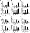
Expression levels of α7 nAChR mRNA and protein in SH-SY5Y cells in which α7 nAChR is overexpressed or silenced. (A) mRNA and (B) protein expression levels of α7 nAChR in SH-SY5Y cells overexpressing α7 nAChR. (C) mRNA and (D) protein expression levels of α7 nAChR in SH-SY5Y cells with silenced α7 nAChR expression. β-actin was used as an internal standard. Expression levels were determined using reverse transcription-quantitative PCR and western blotting. Data are presented as the mean ± standard deviation (n=9).**P<0.01 vs. control. α7 nAChR, nicotinic acetylcholine receptor α7 subunit; shRNA, short hairpin RNA.
Figure 4.
Figure 4.
Expression levels of SYP, PSD-95 and SNAP-25 in SH-SY5Y cells overexpressing α7 nAChR or with silenced α7 nAChR expression. Expression levels of mRNA and protein were determined using reverse transcription-quantitative PCR and western blotting, respectively. mRNA and protein expression levels of (A) SYP, (B) PSD-95 and (C) SNAP-25 after α7 nAChR overexpression. mRNA and protein expression levels of (D) SYP, (E) PSD-95 and (F) SNAP-25 after α7 nAChR silencing. Data are presented as the mean ± standard deviation (n=9). *P<0.05 and **P<0.01 vs. control. SYP, synaptophysin; PSD-95, postsynaptic density of 95 kDa; SNAP-25, synaptosomal-associated protein of 25 kDa; α7 nAChR, nicotinic acetylcholine receptor α7 subunit; shRNA, short hairpin RNA.
Figure 5.
Figure 5.
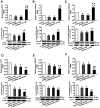
Expression levels of SYP, PSD-95 and SNAP-25 in SH-SY5Y cells following the overexpression of α7 nAChR or silencing of α7 nAChR, and exposure to AβO. mRNA and protein expression levels of (A) SYP, (B) PSD-95 and (C) SNAP-25 after α7 nAChR overexpression. mRNA and protein expression levels of (D) SYP, (E) PSD-95 and (F) SNAP-25 after α7 nAChR silencing. The expression levels of mRNA and protein were determined using reverse transcription-quantitative PCR and western blotting, respectively. Data are presented as the mean ± standard deviation (n=9). *P<0.05 and **P<0.01 vs. control; ##P<0.01, #P<0.05 vs. empty plasmid. SYP, synaptophysin; PSD-95, postsynaptic density of 95 kDa; SNAP-25, synaptosomal-associated protein of 25 kDa; α7 nAChR, nicotinic acetylcholine receptor α7 subunit; AβO, Aβ1-42 oligomer; shRNA, short hairpin RNA.
Figure 6.
Figure 6.
Apoptotic rate of SH-SY5Y cells, which are simultaneously exposed to α7 nAChR overexpression or gene silencing and AβO. (A) Apoptotic rate was determined using flow cytometry and (B) data are presented as the mean ± standard deviation (n=9). **P<0.01 vs. control; #P<0.05 vs. control + AβO. α7 nAChR, nicotinic acetylcholine receptor α7 subunit; AβO, Aβ1-42 oligomer; shRNA, short hairpin RNA.

Similar articles

Cited by

References

    1. Bloom GS. Amyloid-β and tau: The trigger and bullet in Alzheimer disease pathogenesis. JAMA Neurol. 2014;71:505–508. doi: 10.1001/jamaneurol.2013.5847. - DOI - PubMed
    1. Amemori T, Jendelova P, Ruzicka J, Urdzikova LM, Sykova E. Alzheimer's disease: Mechanism and approach to cell therapy. Int J Mol Sci. 2015;16:26417–26451. doi: 10.3390/ijms161125961. - DOI - PMC - PubMed
    1. Ferreira ST, Lourenco MV, Oliveira MM, De Felice FG. Soluble amyloid-β oligomers as synaptotoxins leading to cognitive impairment in Alzheimer's disease. Front Cell Neurosci. 2015;9:191. doi: 10.3389/fncel.2015.00191. - DOI - PMC - PubMed
    1. Viola KL, Klein WL. Amyloid β oligomers in Alzheimer's disease pathogenesis, treatment, and diagnosis. Acta Neuropathol. 2015;129:183–206. doi: 10.1007/s00401-015-1386-3. - DOI - PMC - PubMed
    1. Lesné SE, Sherman MA, Grant M, Kuskowski M, Schneider JA, Bennett DA, Ashe KH. Brain amyloid-β oligomers in ageing and Alzheimer's disease. Brain. 2013;136:1383–1398. doi: 10.1093/brain/awt062. - DOI - PMC - PubMed

MeSH terms