Comparative Study
. 2020 Sep;57(3):697-706.
doi: 10.3892/ijo.2020.5085. Epub 2020 Jun 22.

Comparison of the therapeutic effects of lobaplatin and carboplatin on retinoblastoma in vitro and in vivo

Affiliations
Comparative Study

Comparison of the therapeutic effects of lobaplatin and carboplatin on retinoblastoma in vitro and in vivo

Zijun Zhou et al. Int J Oncol. 2020 Sep.

Abstract

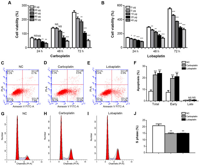
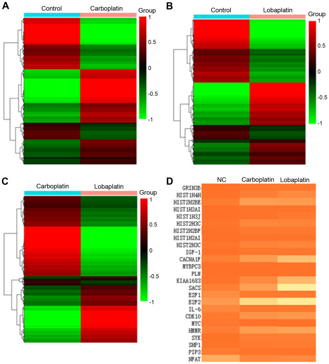
Retinoblastoma (RB) is one of the most aggressive malignancies affecting infants and children. Platinum drugs are commonly used in the treatment of RB; however, their efficacy is often compromised by drug resistance and severe toxicity. The present study aimed to investigate and compare the toxicity and antitumor activity of the third‑generation platinum drugs, carboplatin and lobaplatin, in vitro and in vivo. The Y79 RB cell line was treated with carboplatin or lobaplatin in vitro and then used to establish xenografts in immunodeficient nude mice in vivo; the effects of pharmacological doses of these drugs were then assessed. High concentrations of carboplatin and lobaplatin markedly inhibited Y79 RB cell proliferation in vitro. In addition, the lobaplatin group exhibited higher proportions of early‑stage apoptotic cells than the carboplatin group, while no significant differences in the proportions of cells in the S phase were observed between the 2 groups, as shown by flow cytometry. Significant changes in the E2F1/Cdc25a/Cdk2 pathway in the RB cells were detected by RNA‑seq following carboplatin or lobaplatin intervention. RT‑qPCR, immunofluorescence and immunohistochemical analyses in vivo and in vitro demonstrated that the trends of drug‑induced inhibition of tumor pathological changes may have been regulated through the E2F1/Cdc25a/Cdk2 pathway, and that lobaplatin was more effective than carboplatin in controlling tumors in vivo. On the whole, the findings of the present study demonstrate that lobaplatin is associated with lower cytotoxicity and exerts more prominent therapeutic effects than carboplatin on Y79 RB cells in vitro and in mice in vivo.

Keywords: lobaplatin; carboplatin; retinoblastoma; therapeutic effects.


Figures

Figure 1
Figure 1
Carboplatin and lobaplatin induce various degrees of apoptosis and cell cycle changes in Y79 cells. Cell viability decreased gradually upon treatment with increasing concentrations of (A) carboplatin (0, 20, 40, 60 and 80 µg/ml) or (B) lobaplatin (0, 5, 10, 20 and 40 µg/ml) for 24, 48 or 72 h. The percentages of apoptotic and early apoptotic cells in the carboplatin and lobaplatin groups differed significantly from those in the NC group. (C-F) Representative results of apoptosis in the NC, carboplatin, and lobaplatin groups. (G-J) The proportions of cells in the S phase were lower in the carboplatin and lobaplatin groups than in the NC group. NS, not significant (P>0.05); *P<0.05, **P<0.01 and ***P<0.001 vs. control (no treatment) or NC group.
Figure 2
Figure 2
Gene expression changes in the carboplatin- and lobaplatin-treated groups. (A-C) Heatmaps showing the normalized expression of associated genes in the NC, carboplatin and lobaplatin groups. (D) Results of a cluster analysis of the gene expression levels [in fragments per kilobase per million (FPKM)] in each sample.
Figure 3
Figure 3
Expression of the transcription factors E2F1, Cdc25a and Cdk2 was increased in carboplatin-treated and lobaplatin-treated Y79 cells. Protein and mRNA levels of (A-D) E2F1 (E-H) Cdc25a and (I-L) Cdk2 in Y79 cells detected by RT-PCR analysis and immunofluorescence microscopy in the NC, carboplatin and lobaplatin groups. Magnification, ×40. NS, not significant (P>0.05); ***P<0.001 vs. NC group.
Figure 4
Figure 4
Carboplatin and lobaplatin at pharmacologic doses successfully inhibit the growth of human RB xenografts in vivo. (A) Animal experiments were divided into 4 analyses, including tumor growth curve generation, H&E staining, immunohistochemistry, western blot analysis and RT-qPCR. (B and C) Tumor size in nude mice was monitored after the injection of carboplatin or lobaplatin from day 7 to day 28. H&E staining of the (D-a-g) eyeball and (E-a-g) tumor tissue samples revealed that tumor growth was significantly inhibited after the drug injection, and the degree of inhibition mediated by lobaplatin was greater than that mediated by carboplatin (E-a-g). In (D and E) panels a-g represent the NC, control at 1 week, control at 2 weeks, carboplatin at 1 week, carboplatin at 2 weeks, lobaplatin at 1 week and lobaplatin at 2 weeks, respectively. Magnification, ×1 for tumor size; ×20 for subcutaneous tissue H&E staining and ×2 for eyeball H&E staining. H&E, hematoxylin and eosin.
Figure 5
Figure 5
E2F1, Cdc25a and Cdk2 expression is decreased following carboplatin or lobaplatin treatment in vivo. RT-qPCR was employed to determine the mRNA levels of (A) E2F1, (B) Cdc25a and (C) Cdk2 in the NC, carboplatin and lobaplatin groups two weeks after the mice were treated. Immunohistochemical staining of tumor tissue samples revealed marked decreases in (D-a-g) E2F1-, (E-a-g) Cdc25a- and (F-a-g) Cdk2-positive cell frequencies; panels a-g represent the NC, control at 1 week, control at 2 weeks, carboplatin at 1 week, carboplatin at 2 weeks, lobaplatin at 1 week and lobaplatin at 2 weeks, respectively. Magnification, ×20. NS, not significant (P>0.05); **P<0.01 and ***P<0.001 vs. NC group.

Similar articles

Cited by

References

    1. Hayden B, Jockovich ME, Murray TG, Kralinger MT, Voigt M, Hernandez E, Feuer W, Parel JM. Iontophoretic delivery of carboplatin in a murine model of retinoblastoma. Invest Ophthalmol Vis Sci. 2006;47:3717–3721. doi: 10.1167/iovs.06-0365. - DOI - PubMed
    1. Castillo BV, Jr, Kaufman L. Pediatric tumors of the eye and orbit. Pediatr Clin North Am. 2003;50:149–172. doi: 10.1016/S0031-3955(02)00115-3. - DOI - PubMed
    1. Wiman KG. The retinoblastoma gene: Role in cell cycle control and cell differentiation. FASEB J. 1993;7:841–845. doi: 10.1096/fasebj.7.10.8393817. - DOI - PubMed
    1. MacCarthy A, Birch JM, Draper GJ, Hungerford JL, Kingston JE, Kroll ME, Onadim Z, Stiller CA, Vincent TJ, Murphy MF. Retinoblastoma in Great Britain 1963-2002. Br J Ophthalmol. 2009;93:33–37. doi: 10.1136/bjo.2008.139618. - DOI - PubMed
    1. Shields CL, Mashayekhi A, Au AK, Czyz C, Leahey A, Meadows AT, Shields JA. The International Classification of Retinoblastoma predicts chemoreduction success. Ophthalmology. 2006;113:2276–2280. doi: 10.1016/j.ophtha.2006.06.018. - DOI - PubMed

Publication types

MeSH terms

LinkOut - more resources