. 2020 Sep;46(3):965-976.
doi: 10.3892/ijmm.2020.4651. Epub 2020 Jun 19.

Downregulation of microRNA‑143 promotes osteogenic differentiation of human adipose‑derived mesenchymal stem cells through the k‑Ras/MEK/ERK signaling pathway

Affiliations

Downregulation of microRNA‑143 promotes osteogenic differentiation of human adipose‑derived mesenchymal stem cells through the k‑Ras/MEK/ERK signaling pathway

Yiming Zhang et al. Int J Mol Med. 2020 Sep.

Abstract

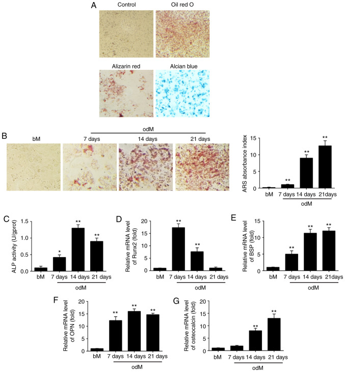
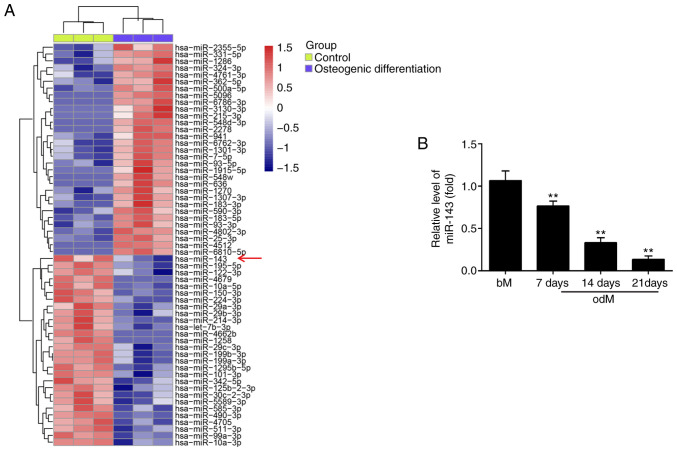
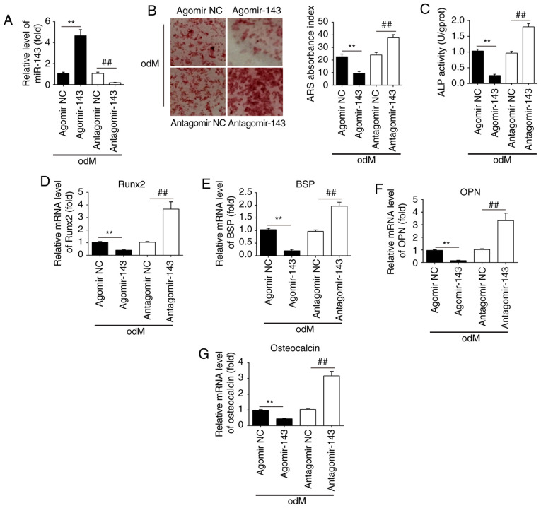
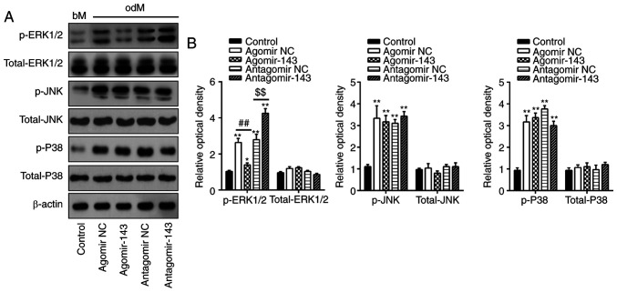
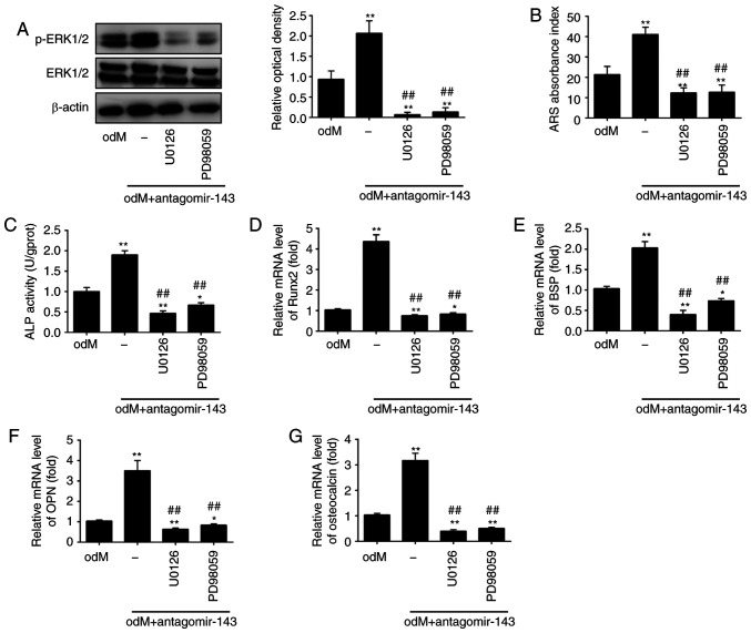
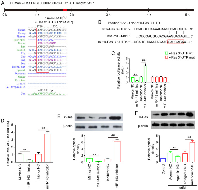
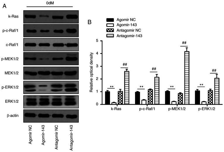
MicroRNAs (miRNAs) are known to have regulatory roles in the osteogenic differentiation of various mesenchymal stem cells (MSCs), although their regulatory role on human adipose‑derived mesenchymal stem cells (hADSCs) remains unclear. The aim of the present study was to investigate the biological function and underlying molecular mechanism of miRNAs in regulating the osteogenic differentiation of hADSCs using microarray assay. hADSCs differentiated into osteoblasts under culture with osteogenic medium, with an increase observed in calcium deposits and alkaline phosphatase activity. The mRNA levels of bone sialoprotein, osteopontin and osteocalcin increased, whereas Runt‑related transcription factor‑2 expression decreased during osteogenic differentiation. In addition, miR‑143 was markedly downregulated during osteogenic differentiation, while miR‑143 overexpression inhibited and miR‑143 knockdown enhanced this process. miR‑143 overexpression also blocked extracellular signal‑regulated kinase 1/2 (ERK1/2) pathway activation, while miR‑143 inhibition enhanced it. The promoting effects of miR‑143 knockdown on the osteogenic differentiation of hADSCs were partly diminished by the mitogen‑activated protein kinase (MEK) inhibitors U0126 and PD98059. Bioinformatics analysis further revealed that miR‑143 targets k‑Ras and directly binds to the 3'‑untranslated region of its mRNA. Inhibition of miR‑143 enhanced the activation of the k‑Ras/MEK/ERK pathway during osteogenic differentiation, whereas miR‑143 overexpression had the opposite effect. Collectively, these results demonstrated that miR‑143 negatively regulates the osteogenic differentiation of hADSCs through the k‑Ras/MEK/ERK pathway, providing further insight into the underlying molecular mechanisms.


Figures

Figure 1
Figure 1
miR-143 was downregulated during hADSC differentiation. (A) hADSCs were seeded in 6-well plates, incubated in normal, adipogenic, osteogenic or chondrogenic differentiation medium for 21 days, and then stained with Oil Red O, Alizarin Red S or Alcian Blue, respectively. Representative images are shown. The control group was stained by Alizarin Red S. Magnification, ×200. (B) Alizarin Red S staining to examine the mineralization of hADSCs cultured in odM or bM at 7, 14 and 21 days. A representative example of three independent experiments is shown. Magnification, ×200. (C) ALP activity was assessed by colorimetric assay. (D) Runx2, (E) BSP, (F) OPN and (G) OCN mRNA levels were assessed by RT-qPCR analysis on days 7, 14 and 21 post-induction of osteogenic differentiation. Data are represented as the mean ± standard deviation of three independent experiments. *P<0.05, **P<0.01 vs. bM group. miR, microRNA; hADSCs, human adipose-derived mesenchymal stem cells; odM, osteogenic differentiation medium; bM, basal medium; ALP, alkaline phosphatase; Runx2, Runt-related transcription factor-2; BSP, bone sialoprotein; OPN, osteopontin; OCN, osteocalcin; RT-qPCR, reverse transcription-quantitative polymerase chain reaction.
Figure 2
Figure 2
miR-143 expression is downregulated during the osteogenic differentiation of hADSCs. (A) Heat map of differentially expressed miRNAs between differentiated and non-differentiated hADSCs. Green indicates low expression levels, and red indicates high expression levels. (B) RT-qPCR analysis was used to assess the expression levels of miR-143 on days 7, 14 and 21 post-induction of osteogenic differentiation. Data are represented as the mean ± standard deviation of three independent experiments. **P<0.01 vs. bM group. miR, microRNA; hADSCs, human adipose-derived mesenchymal stem cells; odM, osteogenic differentiation medium; bM, basal medium; RT-qPCR, reverse transcription-quantitative polymerase chain reaction.
Figure 3
Figure 3
miR-143 negatively regulated the osteogenic differentiation of hADSCs. hADSCs were transfected with agomir-143, antagomir-143 or the corresponding NC (100 nM) for 16 h. The medium was then replaced with odM, and hADSCs were continuously cultured for 14 days. (A) miR-143 expression was assessed by RT-qPCR. (B) Osteogenic differentiation was determined by Alizarin Red S staining and (C) ALP activity was assessed by colorimetric assay on day 14 post-induction. Magnification, ×200. (D) Runx2, (E) BSP, (F) OPN and (G) osteocalcin mRNA levels were assessed by RT-qPCR analysis on day 14 post-induction. Data represent the mean ± standard deviation of three independent experiments. **P<0.01 and ##P<0.01. miR, microRNA; hADSCs, human adipose-derived mesenchymal stem cells; NC, negative control; odM, osteogenic differentiation medium; ALP, alkaline phosphatase; Runx2, Runt-related transcription factor-2; BSP, bone sialoprotein; OPN, osteopontin; RT-qPCR, reverse transcription-quantitative polymerase chain reaction.
Figure 4
Figure 4
miR-143 negatively regulated the ERK1/2 signaling pathway. hADSCs were transfected with agomir-143, antagomir-143 or the corresponding NC (100 nM) for 16 h. The medium was replaced with osteogenic medium, and hADSCs were continuously cultured for 14 days. (A) Western blot analysis was used to assess the protein expression levels of p-ERK1/2, ERK1/2, p-JNK, JNK, p-P38 and P38, with β-actin used as an internal control. (B) The bands were semi-quantitatively analyzed using ImageJ software and normalized to β-actin density. Data represent the mean ± standard deviation of three independent experiments. *P<0.05 and **P<0.01, vs. control group; ##P<0.01 and $$P<0.01. miR, microRNA; hADSCs, human adipose-derived mesenchymal stem cells; NC, negative control; ERK1/2, extracellular-signal regulated kinase 1/2; JNK, c-Jun N-terminal kinase.
Figure 5
Figure 5
miR-143 downregulation enhanced the osteogenic differentiation of human adipose-derived mesenchymal stem cells through activation of the ERK1/2 signaling pathway. Cells were treated with antagomir-143 or antagomir NC, with or without 10 µmol/l U0126 or PD98059 for 15 min. (A) Protein expression levels of p-ERK1/2 and ERK1/2, detected by western blot analysis and quantified using ImageJ software. (B) Graphical representation of the optical density following Alizarin Red S staining at day 21 of culture. (C) ALP activity assessed by colorimetric assay. (D) Runx2, (E) BSP, (F) OPN and (G) osteo-calcin mRNA levels were assessed by reverse transcription-quantitative polymerase chain reaction analysis. Data represent the mean ± standard deviation of three independent experiments. *P<0.05 and **P<0.01, vs. odM group; ##P<0.01 vs. antagomir-143 + odM group. miR, microRNA; ERK1/2, extracellular-signal regulated kinase 1/2; NC, negative control; ALP, alkaline phosphatase; Runx2, Runt-related transcription factor-2; BSP, bone sialoprotein; OPN, osteopontin; odM, osteogenic differentiation medium.
Figure 6
Figure 6
k-Ras was a direct target of miR-143. (A) Sequences of miR-143 binding sites are highly conserved among different species. (B) Putative binding site of miR-143 and k-Ras. (C) Luciferase activity of hADSCs co-transfected with luciferase reporter constructs containing WT or Mut k-Ras 3′-UTR, and with miR-143 mimics, miR-143 inhibitor or NC (n=3). (D) Reverse transcription-quantitative polymerase chain reaction and (E) western blot analysis of mRNA and protein expression levels of k-Ras in hADSCs transfected with miR-143 mimics, miR-143 inhibitor or NC miRNA (n=3). (F) Western blot analysis of k-Ras protein expression in hADSCs transfected with agomir-143, antagomir-143 and the corresponding NC. The medium was replaced with odM after 16 h, and hADSCs were continuously cultured for 14 days prior to protein level assessment. Data represent the mean ± standard deviation of three independent experiments. **P<0.01 and ##P<0.01. miR, microRNA; hADSCs, human adipose-derived mesenchymal stem cells; WT, wild-type; Mut, mutant; NC, negative control; odM, osteogenic differentiation medium.
Figure 7
Figure 7
miR-143 negatively regulated k-Ras/MEK/ERK signaling pathway during osteogenic differentiation of hADSCs. hADSCs were transfected with agomir-143, antagomir-143 or the corresponding NC, and osteogenic differentiation medium was added. (A) Western blot analysis was used to assess the protein expression levels of k-Ras, p-Raf/1, Raf/1, p-MEK1/2, MEK1/2, p-ERK1/2 and ERK1/2, with β-actin used as an internal control. (B) The bands were semi-quantitatively analyzed using ImageJ software and normalized to β-actin density. Data represent the mean ± standard deviation of three independent experiments. **P<0.01 and ##P<0.01. miR, microRNA; hADSCs, human adipose-derived mesenchymal stem cells; MEK, mitogen-activated protein kinase; ERK, extracellular-signal regulated kinase; NC, negative control.

Similar articles

Cited by

References

    1. Locke M, Feisst V, Dunbar PR. Concise review: Human adipose-derived stem cells: Separating promise from clinical need. Stem Cells. 2011;29:404–411. doi: 10.1002/stem.593. - DOI - PubMed
    1. Wang S, Qu X, Zhao RC. Mesenchymal stem cells hold promise for regenerative medicine. Front Med. 2011;5:372–378. doi: 10.1007/s11684-011-0164-4. - DOI - PubMed
    1. Gimble JM, Guilak F. Differentiation potential of adipose derived adult stem (ADAS) cells. Curr Top Dev Biol. 2003;58:137–160. doi: 10.1016/S0070-2153(03)58005-X. - DOI - PubMed
    1. Alvira-González J, Sánchez-Garcés MÀ, Cairó JR, Del Pozo MR, Sánchez CM, Gay-Escoda C. Assessment of bone regeneration using adipose-derived stem cells in critical-size alveolar ridge defects: An experimental study in a dog model. Int J Oral Maxillofac Implants. 2016;31:196–203. doi: 10.11607/jomi.4190. - DOI - PubMed
    1. Sánchez-Garcés MÀ, Alvira-González J, Sánchez CM, Barbany Cairó JR, Del Pozo MR, Gay-Escoda C. Bone regeneration using adipose-derived stem cells with fibronectin in dehiscence-type defects associated with dental implants: An experimental study in a dog model. Int J Oral Maxillofac Implants. 2017;32:e97–e106. doi: 10.11607/jomi.5169. - DOI - PubMed

MeSH terms

Substances