Case Reports
. 2020 Nov;267(11):3157-3160.
doi: 10.1007/s00415-020-10001-7. Epub 2020 Jun 24.

Reversible Encephalopathy Syndrome (PRES) in a COVID-19 patient

Affiliations
Case Reports

Reversible Encephalopathy Syndrome (PRES) in a COVID-19 patient

Lucia Princiotta Cariddi et al. J Neurol. 2020 Nov.

Abstract

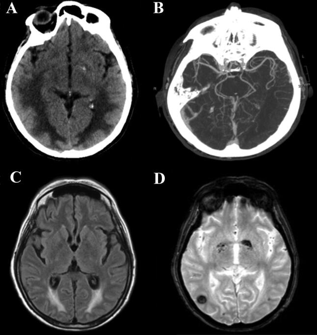
Recently WHO has declared novel coronavirus disease 2019 (COVID-19) outbreak a pandemic. Acute respiratory syndrome seems to be the most common manifestation of COVID-19. Besides pneumonia, it has been demonstrated that SARS-CoV-2 infection affects multiple organs, including brain tissues, causing different neurological manifestations, especially acute cerebrovascular disease (ischemic and hemorrhagic stroke), impaired consciousness and skeletal muscle injury. To our knowledge, among neurological disorders associated with SARS-CoV2 infection, no Posterior Reversible Encephalopathy Syndrome (PRES) has been described yet. Herein, we report a case of a 64-year old woman with COVID19 infection who developed a PRES, and we suggest that it could be explained by the disruption of the blood brain barrier induced by the cerebrovascular endothelial dysfunction caused by SARS-CoV-2.

Keywords: COVID-19; Endothelial dysfunction; Reversible encephalopathy syndrome PRES.


Conflict of interest statement

The authors declare no potential conflicts of interest with respect to the research, authorship, and/or publication of this article.

Figures

Fig.1
Fig.1
Radiological findings. a Brain axial CT on day 25 shows posterior frontal and temporo-parieto-occipital symmetric bilateral hypodensity of the subcortical white matter, and a tiny left occipital parenchymal hemorrhage. b Para-axial CTA scan confirms the absence of vascular malformation and alterations of posterior circle vessel caliber, suggestive of vasoconstriction mechanism. c Axial T2 Flair image on day 56 shows that vasogenic edema is reduced but still detectable and d T2 Gradient-Echo reveals the onset of right temporal hypodensity, correlated to hemorrhagic process

References

    1. Hinchey J, Chaves C, Appignani B, Breen J, Pao L, Wang A, Pessin MS, Lamy C, Mas JL, Caplan LR. A reversible posterior leukoencephalopathy syndrome. N Engl J Med. 1996;334(8):494–500. doi: 10.1056/NEJM199602223340803. - DOI - PubMed
    1. Bartynski WS, Boardman JF, Zeigler ZR, Shadduck RK, Lister J. Posterior reversible encephalopathy syndrome in infection, sepsis, and shock. AJNR Am J Neuroradiol. 2006;27(10):2179–2190. - PMC - PubMed
    1. Racchiusa S, Mormina E, Ax A, Musumeci O, Longo M, Granata F. Posterior reversible encephalopathy syndrome (PRES) and infection: a systematic review of the literature. Neurol Sci. 2019;40(5):915–922. doi: 10.1007/s10072-018-3651-4. - DOI - PubMed
    1. Toledano M, Fugate JE. Posterior reversible encephalopathy in the intensive care unit. Handb Clin Neurol. 2017;141:467–483. doi: 10.1016/B978-0-444-63599-0.00026-0. - DOI - PubMed
    1. Pilato F, Distefano M, Calandrelli R. Posterior reversible encephalopathy syndrome and reversible cerebral vasoconstriction syndrome: clinical and radiological considerations. Front Neurol. 2020;11:34. doi: 10.3389/fneur.2020.00034. - DOI - PMC - PubMed

LinkOut - more resources