Comparative Study
. 2020 Aug;21(4):303-321.
doi: 10.1007/s10162-020-00759-y. Epub 2020 Jun 24.

Ototoxicity and Platinum Uptake Following Cyclic Administration of Platinum-Based Chemotherapeutic Agents

Affiliations
Comparative Study

Ototoxicity and Platinum Uptake Following Cyclic Administration of Platinum-Based Chemotherapeutic Agents

Benjamin K Gersten et al. J Assoc Res Otolaryngol. 2020 Aug.

Abstract

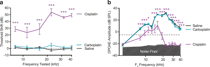
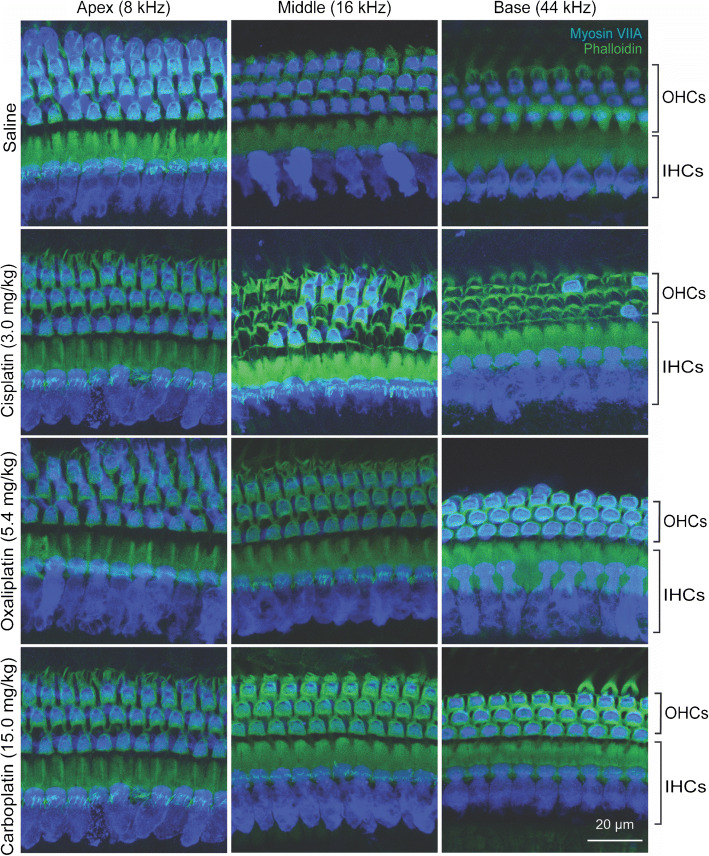
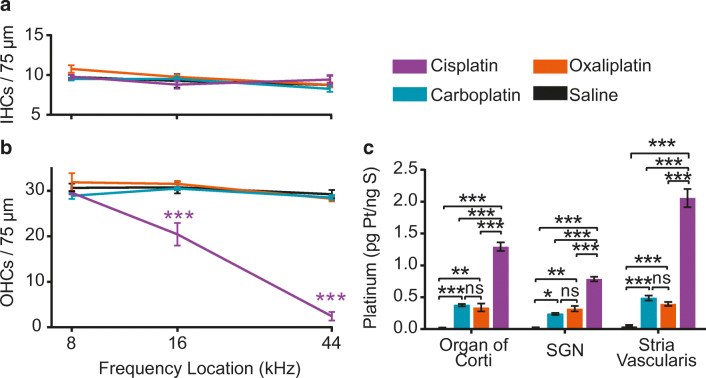
Cisplatin is a widely used anti-cancer drug used to treat a variety of cancer types. One of the side effects of this life-saving drug is irreversible ototoxicity, resulting in permanent hearing loss in many patients. In order to understand why cisplatin is particularly toxic to the inner ear, we compared the hearing loss and cochlear uptake of cisplatin to that of two related drugs, carboplatin and oxaliplatin. These three drugs are similar in that each contains a core platinum atom; however, carboplatin and oxaliplatin are considered less ototoxic than cisplatin. We delivered these three drugs to mice using a 6-week cyclic drug administration protocol. We performed the experiment twice, once using equimolar concentrations of the drugs and once using concentrations of the drugs more proportional to those used in the clinic. For both concentrations, we detected a significant hearing loss caused by cisplatin and no hearing loss caused by carboplatin or oxaliplatin. Cochlear uptake of each drug was measured using inductively coupled plasma mass spectrometry (ICP-MS) to detect platinum. Cochlear platinum levels were highest in mice treated with cisplatin followed by oxaliplatin, while carboplatin was largely excluded from the cochlea. Even when the drug doses were increased, cochlear platinum remained low in mice treated with oxaliplatin or carboplatin. We also examined drug clearance from the inner ear by measuring platinum levels at 1 h and 24 h after drug administration. Our findings suggest that the reduced cochlear platinum we observed with oxaliplatin and carboplatin were not due to increased clearance of these drugs relative to cisplatin. Taken together, our data indicate that the differential ototoxicity among cisplatin, carboplatin, and oxaliplatin is attributable to differences in cochlear uptake of these three drugs.

Keywords: ICP-MS; carboplatin; cisplatin; oxaliplatin.


Figures

Fig. 1
Fig. 1
Cisplatin reduces auditory sensitivity, oxaliplatin and carboplatin do not. Three platinum-based chemotherapeutic drugs were administered to mice in a cyclic drug administration paradigm at equimolar concentrations. a Cisplatin, carboplatin, and oxaliplatin all contain a central platinum atom. b Schematic representation of the experimental protocol including audiometric testing and cyclic drug administration. This figure is adapted from Fernandez et al. . c Threshold shift is reported as the difference in ABR thresholds between pre- and post-drug treatment. Mice that received cisplatin (purple) showed significant threshold shifts at 5 of 6 tested frequencies. Hearing thresholds of carboplatin- (blue) and oxaliplatin-treated (orange) mice were not significantly different from those of saline controls (black). Dashed line at 0 dB represents no change in auditory thresholds relative to baseline. d OHC function was measured using DPOAEs. Relative to saline-treated control mice (black), cisplatin (purple) caused significantly reduced DPOAE amplitudes at 11 out of 14 tested f2 frequencies. Neither carboplatin nor oxaliplatin caused decreased DPOAE amplitudes at any frequency. Gray shaded region indicates the mean biological noise floor measured during recordings. Noise floor measurements were taken and plotted for each treatment condition. Dashed line at – 5 dB SPL represents system noise level below which DPOAEs are considered absent. All data are presented as group means ± SEM. N = 8–10 mice for all groups. *P < 0.05, **P < 0.01, ***P < 0.001 between cisplatin and saline. (ABR: 2-way ANOVA with Sidak’s test for multiple comparisons, F(3, 181) = 116.1, P < 0.001; DPOAE: 2-way ANOVA, F(3, 444) = 68.19, P < 0.001)
Fig. 2
Fig. 2
Cisplatin causes death of outer hair cells (OHCs); oxaliplatin and carboplatin do not. Isolated cochlear turns were stained for myosin-VIIA (blue) and phalloidin (green). Cochlear inner and outer hair cells were imaged at three distinct locations along the basilar membrane. Saline-treated control mice showed no loss of inner or outer hair cells. In cisplatin-treated mice, inner hair cells (IHCs) remained intact while outer hair cells were missing in the middle and basal cochlear turns. Oxaliplatin and carboplatin did not result in inner or outer hair cell loss in any cochlear turn. Scale bar = 20 μm and applies to all panels
Fig. 3
Fig. 3
Cisplatin kills outer hair cells and results in the highest levels of cochlear platinum. Cisplatin was administered at a daily dose of 3 mg/kg, carboplatin was administered at a daily dose of 3.71 mg/kg and oxaliplatin was administered at a daily dose of 3.97 mg/kg. These equimolar doses were chosen so each animal was exposed to the same amount of platinum regardless of the treatment group to which it was assigned. a Inner hair cell counts from mice in all drug administration groups. No significant differences in IHC density were found between saline controls and drug-treated groups at any cochlear location. b Outer hair cell counts from mice in all drug administration groups. At middle and basal cochlear turns, there was significant OHC loss in cisplatin-treated mice compared with saline-treated controls. Cisplatin caused 39 % loss of OHCs in the middle turn and 93 % loss of OHCs in the basal turn of the cochlea. Oxaliplatin and carboplatin treatment did not result in loss of outer hair cells. Data displayed are group means ± SEM. N = 3–7 cochleas per drug condition. 2-way ANOVA with Sidak’s test for multiple comparisons (F(3, 51) = 263.3, P < 0.001).c After three cycles of drug administration, whole cochleas and microdissected cochlear samples were analyzed for platinum content using ICP-MS. Cochleas from cisplatin-treated animals contained significantly increased platinum levels relative to controls in all tissues, including whole cochleas and microdissected organ of Corti, SGN, and stria vascularis, with highest levels in stria vascularis. Tissues from oxaliplatin-treated mice contained significantly increased levels of platinum relative to controls in whole cochlea, organ of Corti, and stria vascularis. In cochleas from carboplatin-treated mice, no significant elevation in platinum was detected in any samples relative to controls. There was a significant difference in platinum accumulation across tissue types (2-way ANOVA with Sidak’s test for multiple comparisons, F(3, 88) = 32.07, P < 0.001) and drug types (2-way ANOVA with Sidak’s test for multiple comparisons, F(3, 88) = 215.6, P < 0.001). Data displayed are group means ± SEM. N = 5–9 samples per condition and tissue type. *P < 0.01, ***P < 0.001
Fig. 4
Fig. 4
Drug clearance from the cochlea does not account for the differences in platinum levels. a In order to determine if the observed differences in cochlear platinum levels were due to differences in drug clearance, we measured cochlear platinum levels 1 h and 24 h after administering the equivalent of one drug injection period (4 days) of equimolar solutions via a single i.p. injection. b Platinum levels were measured in four tissue types treated with either cisplatin, carboplatin, or oxaliplatin and compared relative to saline-treated controls. Platinum levels were significantly dependent on drug type at both 1-h (2-way ANOVA, F(3, 44) = 233.5, P < 0.001) and 24-h following injection (2-way ANOVA, F(3, 44) = 50.66, P < 0.001). Dunnett’s post-hoc test for multiple comparisons revealed significant platinum uptake in all 4 cisplatin-treated tissue types, no carboplatin-treated tissue types, and some oxaliplatin-treated tissue types (spiral ganglion neurons, stria vascularis, and whole cochleas). Solid lines represent platinum levels detected in drug-treated samples; dashed lines represent platinum levels detected in saline-treated control samples. Data displayed are group means ± SEM. N = 3–4 samples per treatment condition and time point. *P < 0.05, **P < 0.01, ***P < 0.001
Fig. 5
Fig. 5
At clinically proportional concentrations, carboplatin does not cause auditory dysfunction. a Following the clinically proportional drug administration protocol, ABR threshold shifts were measured for animals treated with cisplatin (purple), carboplatin (blue) delivered at a dose 5 times greater than cisplatin, or saline controls (black). Cisplatin-treated animals showed increased threshold shifts relative to saline-treated controls at all tested frequencies (2-way ANOVA with Sidak’s test for multiple comparisons, F(2, 106) = 220.1, P < 0.001). Carboplatin treatment did not result in threshold shifts at any tested frequency despite the higher dose of carboplatin. b DPOAEs were measured as an indirect assessment of OHC function. Cisplatin-treated mice had reduced DPOAE amplitudes at 12 of the 14 tested f2 frequencies, and carboplatin-treated animals showed no changes relative to saline-treated controls at any frequency (2-Way ANOVA with Sidak’s test for multiple comparisons, F(2,252) = 382.8, P < 0.0001). Data displayed are group means ± SEM. N = 6–8 subjects per treatment condition. *P < 0.05, **P < 0.01, *** P < 0.001
Fig. 6
Fig. 6
At clinically proportional doses, only cisplatin causes OHC loss. Isolated cochlear turns were stained for myosin-VIIA (blue) and phalloidin (green). Cochlear inner and outer hair cells were imaged and counted at three distinct locations along the basilar membrane. Control (saline-treated) mice showed no loss of IHCs or OHCs. Cisplatin-treated mice showed no loss of IHCs and significant loss of OHCs in the basal and middle cochlear turns. Oxaliplatin and carboplatin did not result in IHC or OHC loss in any cochlear turn. Scale bar = 20 μm and applies to all panels
Fig. 7
Fig. 7
At clinically proportional doses, neither carboplatin nor oxaliplatin caused a decrease in HC density, and cochlear platinum levels following carboplatin or oxaliplatin treatment remain lower than those of cisplatin. The cumulative dose of cisplatin remained unchanged relative to the equimolar experiment at 36 mg/kg. Carboplatin was administered at 5 times that dose at 180 mg/kg. Oxaliplatin was administered at 1.2 times the cisplatin dose at 43.2 mg/kg. a No significant differences in IHC density were found between saline-treated controls and drug-treated groups at any cochlear location. b OHC density was significantly dependent on drug treatment (2-way ANOVA, F(3, 76) = 73.3, P < 0.001). Cisplatin-treated mice showed significant OHC loss at middle (34 %) and basal (92 %) cochlear turns compared with saline controls (Sidak’s test for multiple comparisons, P < 0.001). Oxaliplatin or carboplatin treatment did not result in significant loss of OHCs at any cochlear location. Data displayed are group means ± SEM. N = 4–8 cochleas per condition. ***P < 0.001. c Microdissected cochlear samples were assessed for platinum content using ICP-MS. Cochlear platinum levels were significantly elevated for all samples treated with platinum-containing drugs relative to saline controls (2-way ANOVA with Sidak’s test for multiple comparisons, F(3, 72) = 358.2, P < 0.001). Cochlear platinum levels following carboplatin and oxaliplatin were significantly lower than those following cisplatin. Data presented are mean ± SEM. N = 5–10 samples per condition. *P < 0.05, **P < 0.01, ***P < 0.001

Similar articles

Cited by

References

    1. Al-Batran SE, Hartmann JT, Probst S, Schmalenberg H, Hollerbach S, Hofheinz R, Rethwisch V, Seipelt G, Homann N, Wilhelm G, Schuch G, Stoehlmacher J, Derigs HG, Hegewisch-Becker S, Grossmann J, Pauligk C, Atmaca A, Bokemeyer C, Knuth A, Jager E. Phase III trial in metastatic gastroesophageal adenocarcinoma with fluorouracil, leucovorin plus either oxaliplatin or cisplatin: a study of the Arbeitsgemeinschaft Internistische Onkologie. J Clin Oncol. 2008;26:1435–1442. - PubMed
    1. Andersson A, Fagerberg J, Lewensohn R, Ehrsson H. Pharmacokinetics of cisplatin and its monohydrated complex in humans. J Pharm Sci. 1996;85:824–827. - PubMed
    1. Astolfi L, Simoni E, Ciorba A, Martini A. In vitro protective effects of Ginkgo bilboa against cisplatin toxicity in mouse cell line OCk3. Audiol Med. 2008;6(4):251–258.
    1. Bass JK, Huang J, Onar-Thomas A, Chang KW, Bhagat SP, Chintagumpala M, Bartels U, Gururangan S, Hassall T, Heath JA, McCowage G, Cohn RJ, Fisher MJ, Robinson G, Broniscer A, Gajjar A, Gurney JG. Concordance between the Chang and the International Society of Pediatric Oncology (SIOP) ototoxicity grading scales in patients treated with cisplatin for medulloblastoma. Pediatr Blood Cancer. 2014;61:601–605. - PMC - PubMed
    1. Becouarn Y, Agostini C, Trufflandier N, Boulanger V. Oxaliplatin: available data in non-colorectal gastrointestinal malignancies. Crit Rev Oncol Hematol. 2001;40:265–272. - PubMed

Publication types

LinkOut - more resources