Review
. 2020 Aug;24(15):8276-8290.
doi: 10.1111/jcmm.15301. Epub 2020 Jun 25.

Lysosomal dysfunction-induced autophagic stress in diabetic kidney disease

Affiliations
Review

Lysosomal dysfunction-induced autophagic stress in diabetic kidney disease

Hui Juan Zheng et al. J Cell Mol Med. 2020 Aug.

Abstract

The catabolic process that delivers cytoplasmic constituents to the lysosome for degradation, known as autophagy, is thought to act as a cytoprotective mechanism in response to stress or as a pathogenic process contributing towards cell death. Animal and human studies have shown that autophagy is substantially dysregulated in renal cells in diabetes, suggesting that activating autophagy could be a therapeutic intervention. However, under prolonged hyperglycaemia with impaired lysosome function, increased autophagy induction that exceeds the degradative capacity in cells could contribute toward autophagic stress or even the stagnation of autophagy, leading to renal cytotoxicity. Since lysosomal function is likely key to linking the dual cytoprotective and cytotoxic actions of autophagy, it is important to develop novel pharmacological agents that improve lysosomal function and restore autophagic flux. In this review, we first provide an overview of the autophagic-lysosomal pathway, particularly focusing on stages of lysosomal degradation during autophagy. Then, we discuss the role of adaptive autophagy and autophagic stress based on lysosomal function. More importantly, we focus on the role of autophagic stress induced by lysosomal dysfunction according to the pathogenic factors (including high glucose, advanced glycation end products (AGEs), urinary protein, excessive reactive oxygen species (ROS) and lipid overload) in diabetic kidney disease (DKD), respectively. Finally, therapeutic possibilities aimed at lysosomal restoration in DKD are introduced.

Keywords: autophagic stress; autophagy; diabetic kidney disease; lysosomal dysfunction.


Conflict of interest statement

All authors declare that they have no conflicts of interest.

Figures

FIGURE 1
FIGURE 1
Schematic depiction of the autophagic‐lysosome pathway. Protein aggregates and damaged mitochondria are selectively targeted for autophagy via ubiquitin (Ub)‐labelled adaptor proteins (eg p62, Nix and NBR1). Autophagy occurs when the autophagy receptors bind cargoes with LC3 to initiate substrate sequestration in the forming autophagosome. Matured autophagosomes fuse with lysosomes containing hydrolytic enzymes for cargo degradation. When the degradation is complete inside the autolysosome, the lysosomes regain their original properties via lysosome re‐formation. The lysosome comprises a specific set of luminal and integral‐membrane proteins. The acidification of the lysosome is maintained by the vacuolar‐H+ ATPase and the lysosomal membrane, which contains highly glycosylated lysosome‐associated membrane proteins (LAMPs), ion channels and transporters, and cholesterol transporters. Moreover, the biogenesis and function of lysosomes are also regulated by transcription factors such as TFEB. As terminal degradation stations, normal morphology and function of lysosomes are essential for successful completion of autophagic degradation
FIGURE 2
FIGURE 2
Schematic diagram displaying the dual roles of autophagy based on lysosomal function in kidney. When lysosome function is normal, proper autophagy induction in response to stress factors maintains or restores cellular homeostasis to protect against cell injury and thus promotes cell survival in the kidney. Prolonged and intense autophagy induction in response to damaged cellular constituents exceeding the degradative capacity of lysosomes or normal autophagy induction accompanied by marked impairment of lysosomal degradation, result in autophagic stress. The condition of autophagic stress contributes to aggravated cell damage and/or autophagic cell death in kidneys
FIGURE 3
FIGURE 3
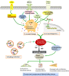
Schematic diagram displaying the role of autophagic stress in various pathogenic factors for diabetic kidney disease. In a chronic diabetic state, various pathogenic factors of diabetic kidney disease, such as prolonged hyperglycaemia, advanced glycosylation end products (AGEs) overload, urinary protein overload, lipid droplets accumulation and excessive reactive oxygen species (ROS), trigger lysosomal dysfunction due to lysosomal membrane permeabilization (LMP) and/or reduced lysosomal biogenesis. Under these conditions, increased or sustained demand for autophagy induction in cells accompanied by the impaired clearance of cellular components promotes autophagic stress, resulting in the accumulation of damaged organelles and/or abnormal proteins in renal cells. The condition of autophagic stress in renal cells causes a switch from a renoprotective mechanism to a cytotoxic state, which is implicated in the pathogenesis of diabetic kidney disease

Similar articles

Cited by

References

    1. Zhang L, Long J, Jiang W, et al. Trends in chronic kidney disease in China. N Engl J Med. 2016;375:905‐906. - PubMed
    1. Thomas MC, Brownlee M, Susztak K, et al. Diabetic kidney disease. Nat Rev Dis Primers. 2015;1:15018. - PMC - PubMed
    1. Jefferson JA, Shankland SJ, Pichler RH. Proteinuria in diabetic kidney disease: a mechanistic viewpoint. Kidney Int. 2008;74:22‐36. - PubMed
    1. Warren AM, Knudsen ST, Cooper ME. Diabetic nephropathy: an insight into molecular mechanisms and emerging therapies. Expert Opin Ther Targets. 2019;23:579‐591. - PubMed
    1. Jha JC, Banal C, Chow BS, et al. Diabetes and kidney disease: role of oxidative stress. Antioxid Redox Signal. 2016;25:657‐684. - PMC - PubMed

Publication types

Substances