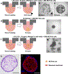
Cancer Cell Invasion of Mammary Organoids with Basal-In Phenotype
- PMID: 32583612
- PMCID: PMC7759600
- DOI: 10.1002/adhm.202000810
Cancer Cell Invasion of Mammary Organoids with Basal-In Phenotype
Abstract
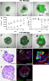
This paper describes mammary organoids with a basal-in phenotype where the basement membrane is located on the interior surface of the organoid. A key materials consideration to induce this basal-in phenotype is the use of a minimal gel scaffold that the epithelial cells self-assemble around and encapsulate. When MDA-MB-231 breast cancer cells are co-cultured with epithelial cells from day 0 under these conditions, cells self-organize into patterns with distinct cancer cell populations both inside and at the periphery of the epithelial organoid. In another type of experiment, the robust formation of the basement membrane on the epithelial organoid interior enables convenient studies of MDA-MB-231 invasion in a tumor progression-relevant direction relative to epithelial cell-basement membrane positioning. That is, the study of cancer invasion through the epithelium first, followed by the basement membrane to the basal side, is realized in an experimentally convenient manner where the cancer cells are simply seeded on the outside of preformed organoids, and their invasion into the organoid is monitored. Interestingly, invasion is more prominent when tumor cells are added to day 7 organoids with less developed basement membranes compared to day 16 organoids with more defined ones.
Keywords: basal-in phenotype; basement membrane; breast cancer; invasion; organoid.
© 2020 WILEY-VCH Verlag GmbH & Co. KGaA, Weinheim.
Figures


Similar articles
-
Triple-negative breast cancer cells invade adipocyte/preadipocyte-encapsulating geometrically inverted mammary organoids.Integr Biol (Camb). 2023 Apr 11;15:zyad004. doi: 10.1093/intbio/zyad004. Integr Biol (Camb). 2023. PMID: 37015816 Free PMC article.
-
Basement membrane formation of fetal mouse intestinal epithelial cells in organoid cultures.Acta Anat (Basel). 1995;153(2):96-105. doi: 10.1159/000147719. Acta Anat (Basel). 1995. PMID: 8560971
-
Mammary organoids from immature virgin rats undergo ductal and alveolar morphogenesis when grown within a reconstituted basement membrane.Exp Cell Res. 1991 Sep;196(1):49-65. doi: 10.1016/0014-4827(91)90455-4. Exp Cell Res. 1991. PMID: 1879471
-
Matrix scaffolds for endometrium-derived organoid models.Front Endocrinol (Lausanne). 2023 Aug 10;14:1240064. doi: 10.3389/fendo.2023.1240064. eCollection 2023. Front Endocrinol (Lausanne). 2023. PMID: 37635971 Free PMC article. Review.
-
The matrix environmental and cell mechanical properties regulate cell migration and contribute to the invasive phenotype of cancer cells.Rep Prog Phys. 2019 Jun;82(6):064602. doi: 10.1088/1361-6633/ab1628. Epub 2019 Apr 4. Rep Prog Phys. 2019. PMID: 30947151 Review.
Cited by
-
Triple-negative breast cancer cells invade adipocyte/preadipocyte-encapsulating geometrically inverted mammary organoids.Integr Biol (Camb). 2023 Apr 11;15:zyad004. doi: 10.1093/intbio/zyad004. Integr Biol (Camb). 2023. PMID: 37015816 Free PMC article.
-
Harnessing the power of artificial intelligence for human living organoid research.Bioact Mater. 2024 Aug 30;42:140-164. doi: 10.1016/j.bioactmat.2024.08.027. eCollection 2024 Dec. Bioact Mater. 2024. PMID: 39280585 Free PMC article. Review.
-
Programming temporal stiffness cues within extracellular matrix hydrogels for modelling cancer niches.Mater Today Bio. 2024 Feb 16;25:101004. doi: 10.1016/j.mtbio.2024.101004. eCollection 2024 Apr. Mater Today Bio. 2024. PMID: 38420142 Free PMC article.
-
In Vitro Organoid-Based Assays Reveal SMAD4 Tumor-Suppressive Mechanisms for Serrated Colorectal Cancer Invasion.Cancers (Basel). 2023 Dec 13;15(24):5820. doi: 10.3390/cancers15245820. Cancers (Basel). 2023. PMID: 38136364 Free PMC article.
-
Development of robust antiviral assays using relevant apical-out human airway organoids.bioRxiv [Preprint]. 2024 Jul 12:2024.01.02.573939. doi: 10.1101/2024.01.02.573939. bioRxiv. 2024. PMID: 38260306 Free PMC article. Preprint.
References
-
- Saglam-Metiner P, Gulce-Iz S, Biray-Avci C, Gene 2019, 686, 203. - PubMed
-
- Sachs N, de Ligt J, Kopper O, Gogola E, Bounova G, Weeber F, Balgobind AV, Wind K, Gracanin A, Begthel H, Korving J, van Boxtel R, Duarte AA, Lelieveld D, van Hoeck A, Ernst RF, Blokzijl F, Nijman IJ, Hoogstraat M, van de Ven M, Egan DA, Zinzalla V, Moll J, Boj SF, Voest EE, Wessels L, van Diest PJ, Rottenberg S, Vries RGJ, Cuppen E, Clevers H, Cell 2018, 172, 373. - PubMed
-
- Huch M, Knoblich JA, Lutolf MP, Martinez-Arias A, Dev. 2017, 144, 938. - PubMed
Publication types
MeSH terms
Grants and funding
LinkOut - more resources
Full Text Sources
Other Literature Sources
Miscellaneous

