. 2020 Aug 15;31(18):1963-1973.
doi: 10.1091/mbc.E20-02-0100. Epub 2020 Jun 17.

SCYL1 arginine methylation by PRMT1 is essential for neurite outgrowth via Golgi morphogenesis

Affiliations

SCYL1 arginine methylation by PRMT1 is essential for neurite outgrowth via Golgi morphogenesis

Genki Amano et al. Mol Biol Cell. .

Abstract

Arginine methylation is a common posttranslational modification that modulates protein function. SCY1-like pseudokinase 1 (SCYL1) is crucial for neuronal functions and interacts with γ2-COP to form coat protein complex I (COPI) vesicles that regulate Golgi morphology. However, the molecular mechanism by which SCYL1 is regulated remains unclear. Here, we report that the γ2-COP-binding site of SCYL1 is arginine-methylated by protein arginine methyltransferase 1 (PRMT1) and that SCYL1 arginine methylation is important for the interaction of SCYL1 with γ2-COP. PRMT1 was colocalized with SCYL1 in the Golgi fraction. Inhibition of PRMT1 suppressed axon outgrowth and dendrite complexity via abnormal Golgi morphology. Knockdown of SCYL1 by small interfering RNA (siRNA) inhibited axon outgrowth, and the inhibitory effect was rescued by siRNA-resistant SCYL1, but not SCYL1 mutant, in which the arginine methylation site was replaced. Thus, PRMT1 regulates Golgi morphogenesis via SCYL1 arginine methylation. We propose that SCYL1 arginine methylation by PRMT1 contributes to axon and dendrite morphogenesis in neurons.


Figures

FIGURE 1:
FIGURE 1:
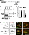
Arginine methylation of SCYL1 is important for the interaction between SCYL1 and γ2-COP. (A) HeLa cells transfected with SCYL1-Flag were treated or not treated with 40 µM AdOx (a methylation inhibitor) for 24 h before immunoprecipitation with an anti-Flag antibody. The immunoprecipitates were subjected to SDS–PAGE and immunoblot analysis with ASYM24 (asymmetrical dimethylated arginine) and anti-Flag and anti-γ2-COP antibodies. (B) Quanti­fication of immunoblots from immunoprecipitation assays with anti-Flag antibody. Values were normalized to controls. Unpaired, two-tailed Student’s t test; **p  <  0.01. Error bars indicate the mean  ±  SEM (n  = 4). (C) HeLa cells expressing SCYL1-Flag were treated or not treated with 40 µM AdOx for 24 h before fixation. The cells were immunostained with anti-γ2-COP (green) and anti-Flag (red) antibodies. The boxed areas (1, 2) are enlarged in the right panels. Results are representative of more than three experiments. Scale bar: 20 μm.
FIGURE 2:
FIGURE 2:
C-terminal arginine methylation of SCYL1 is crucial for the interaction of SCYL1 with γ2-COP. (A) Alignment of the human SCYL1 amino acid sequence. SCYL1 has two isoforms. Both isoforms have an Arg–Lys–Leu–Asp sequence at the C-terminus. aa: amino acid. (B) HeLa cells were transfected with wild-type (RKLD) or mutant (AKLD) SCYL1-Flag for 24 h. Immuno­precipitation assays were performed with an anti-Flag antibody. The immunoprecipitates were subjected to SDS–PAGE and immunoblot analysis with ASYM24 (asymmetrical dimethylated arginine) and with anti-Flag and anti-γ2-COP antibodies. (C) Quantification of immunoblots from immunoprecipitation assays with the anti-Flag antibody. Values were normalized to controls. Unpaired, two-tailed Student’s t test; **p  <  0.01, ***p < 0.001. The data are shown as the mean  ±  SEM (n  = 7). (D) HeLa cells were transfected with wild-type (RKLD) or mutant (AKLD) SCYL1-Flag for 24 h. The cells were fixed and immunostained with anti-γ2-COP (green) and anti-Flag (red) antibodies. The boxed areas (1, 2) are enlarged in the right panels. Results are representative of more than three experiments. Scale bar: 20 μm
FIGURE 3:
FIGURE 3:
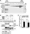
PRMT1 is colocalized with SCYL1 in microsomal fractions and regulates SCYL1 arginine methylation for the interaction between SCYL1 and γ2-COP. (A) Microsomal fractions extracted from HeLa cells were ultracentrifuged using iodixanol concentration gradients. After ultracentrifugation, 12 fractions were collected from the upper layer and immunoblot analysis was performed with anti-GM130 (a cis-Golgi marker), anti-ERGIC53 (an ER-Golgi intermediate compartment marker), anti-Calnexin (an ER-associated protein), anti-ARF4 (a COPI vesicle-associated small GTPase), anti-γ2-COP, anti-SCYL1, and anti-PRMT1 antibodies. (B) HeLa cells were treated with control or PRMT1 siRNA for 24 h before being transfected with SCYL1-Flag. Immunoprecipitation assays were performed with an anti-Flag antibody. The immunoprecipitates were subjected to SDS–PAGE and immunoblot analysis with ASYM24 (asymmetrical dimethylated arginine) and with anti-Flag and anti-γ2-COP antibodies. The asterisk indicates nonspecific bands. (C) Quantification of immunoblots of immunoprecipitates. Values were normalized to controls. Unpaired, two-tailed Student’s t test; *p  <  0.05, **p  <  0.01. Error bars indicate the mean  ±  SEM (n  = 5).
FIGURE 4:
FIGURE 4:
PRMT1 regulates Golgi organization. (A) HeLa cells were transfected with control or PRMT1 siRNA for 24 h. The samples were subjected to SDS–PAGE and immunoblot analysis with anti-PRMT1, anti-GM130 (a cis-Golgi marker), anti-GRP78 (an ER chaperone protein), anti-ERGIC53 (an ER-Golgi intermediate compartment marker), anti-SCYL1, anti-γ2-COP, anti-ARF4 (a COPI vesicle-associated small GTPase), and anti-GAPDH antibodies. The asterisks indicate nonspecific bands. (B) Expression levels of indicated proteins were measured by quantitative immunoblotting. Values were normalized to controls. Unpaired, two-tailed Student’s t test; *p  <  0.05, ***p  <  0.001. Error bars indicate the mean  ±  SEM (n  =  6). ns: not significant. (C) HeLa cells were transfected with control siRNA or PRMT1 siRNA. The cells were immunostained with anti-PRMT1 (green) and anti-GM130 (red) antibodies. The boxed areas (1, 2) are enlarged in the right panels. The arrowheads indicate PRMT1 knockdown cells. Representative results from three independent experiments are shown. Scale bars: 20 μm.
FIGURE 5:
FIGURE 5:
Endogenous SCYL1 is arginine- methylated. (A) HEK293T cells were treated with control or PRMT1 siRNA for 48 h. Endogenous SCYL1 was immunoprecipitated with an anti-SCYL1 antibody. The immunoprecipitates were subjected to SDS–PAGE and immunoblot analysis with ASYM24 and anti-SCYL1 antibodies. The asterisk indicates an immunoglobulin heavy chain. (B) Immunoprecipitation of endogenous SCYL1 from mouse brain lysate. Extract of developmental mouse brain was incubated with rabbit IgG or anti-SCYL1 antibody. The immunoprecipitates were subjected to SDS–PAGE and immunoblot analysis with ASYM24 and anti-SCYL1 antibodies. The asterisk indicates an immunoglobulin heavy chain. (C) Immunoprecipitation of endogenous SCYL1 from cultured neuron lysate. After rat hippocampal neurons were cultured for 3 d, immunoprecipitation assays were performed with rabbit IgG or anti-SCYL1 antibody. The immunoprecipitates were analyzed by immunoblotting with ADMA (asymmetrical dimethylated arginine) and anti-SCYL1 antibodies. The asterisk indicates an immunoglobulin heavy chain.
FIGURE 6:
FIGURE 6:
Arginine methylation is important for axon outgrowth. (A) Hippocampal neurons were treated for 48 h with 10 µM AMI-1 (a PRMT1 inhibitor). The samples were subjected to SDS–PAGE and immunoblot analysis with the indicated antibodies. (B) Expression levels of indicated proteins were measured by quantitative immunoblotting. Values were normalized to controls. Unpaired, two-tailed Student’s t test; ***p  <  0.001. Error bars indicate the mean  ±  SEM (n  =  3). ns: not significant. (C) Hippocampal neurons were cultured in the presence of AMI-1 or AdOx for 48 h before fixation. Neurons were fixed at 3 DIV and then immunostained with Tau-1 antibody, an axonal marker. Scale bar: 100 μm. (D) Axon length was measured at 3 DIV in neurons treated with the indicated inhibitors. Unpaired, two-tailed Student’s t test; ***p  <  0.001. The data are shown as the mean  ±  SEM of three independent experiments (120 axons from each group). (E) Cumulative frequency plots showing the distribution of axon length from individual neurons that were analyzed in D.
FIGURE 7:
FIGURE 7:
Arginine methylation is essential for dendrite complexity. (A) Hippocampal neurons were transfected with GFP at 10 DIV. Neurons were fixed at 14 DIV after treatment with indicated inhibitors for 72 h and then immunostained with an anti-GFP antibody. Scale bar: 100 μm. (B) Illustration of Sholl analysis used to quantify neurite complexity. The number of interactions made by the extending neurites with each circle was counted and used as a measure of neurite complexity. (C) Sholl analysis of neurite complexity in neurons treated as in (A). The mean numbers of intersections are plotted. Unpaired, two-tailed Student’s t test; ***p  <  0.001. Error bars indicate the mean  ±  SEM of four independent experiments (40 hippocampal neurons from each group).
FIGURE 8:
FIGURE 8:
C-terminal arginine methylation of SCYL1 is important for axon outgrowth. (A) Rat-1 cells were transfected with control or SCYL1 siRNA. The samples were subjected to SDS–PAGE and immunoblot analysis with anti-SCYL1 and anti-GAPDH antibodies. (B) Cotransfection of GST and SCYL1 siRNA with siRNA-resistant SCYL1-Flag (wild-type or mutant) was performed in cultured hippocampal neurons by electroporation. Neurons were fixed at 3 DIV and then immunostained with anti-Flag (green) and anti-GST (red) antibodies. Scale bar: 100 μm. (C) Axon length was measured at 3 DIV in neurons treated as in B. Unpaired, two-tailed Student’s t test; ***p < 0.001. ns: not significant. The data are shown as the mean  ±  SEM of 120 axons from each group.
FIGURE 9:
FIGURE 9:
Schematic representation illustrating roles of SCYL1 arginine methylation by PRMT1 in neurons. C-terminal arginine of SCYL1 is methylated by PRMT1. SCYL1 methylated by PRMT1 inter­acts with γ2-COP. COPI vesicles are formed via the interaction between methylated SCYL1 and γ2-COP. Golgi organization by COPI vesicles enables posttranslational modification of axonal and dendritic proteins and vesicle trafficking, thereby enhancing axon elongation and dendrite branching.

References

    1. Bartel RL, Borchardt RT. (1984). Effects of adenosine dialdehyde on S-adenosylhomocysteine hydrolase and S-adenosylmethionine-dependent transmethylations in mouse L929 cells. Mol Pharmacol , 418–424. - PubMed
    1. Bedford MT, Clarke SG. (2009). Protein arginine methylation in mammals: who, what, and why. Mol Cell , 1–13. - PMC - PubMed
    1. Burman JL, Bourbonniere L, Philie J, Stroh T, Dejgaard SY, Presley JF, McPherson PS. (2008). Scyl1, mutated in a recessive form of spinocerebellar neurodegeneration, regulates COPI-mediated retrograde traffic. J Biol Chem , 22774–22786. - PubMed
    1. Burman JL, Hamlin JNR, McPherson PS. (2010). Scyl1 regulates Golgi morphology. PLoS One , e9537. - PMC - PubMed
    1. Chafe SC, Mangroo D. (2010). Scyl1 facilitates nuclear tRNA export in mammalian cells by acting at the nuclear pore complex. Mol Biol Cell , 2483–2499. - PMC - PubMed

Publication types

MeSH terms

LinkOut - more resources